Dog Histiocytoma: 7 Warning Signs, Causes & Treatment Options
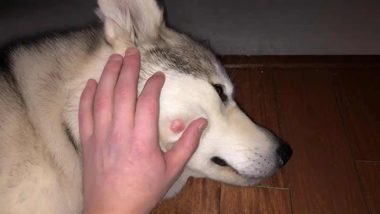
Table of Contents
Dog histiocytoma is a common, often benign, skin growth that frequently causes concern among pet owners due to its sudden appearance and rapid growth. While the word “tumour” can understandably trigger alarm, histiocytomas are typically non-cancerous and usually resolve on their own without aggressive intervention. These distinctive lumps, sometimes referred to as “button tumours”, are a prevalent dermatological issue, particularly in younger canines. Understanding the nature of dog histiocytoma, how it presents, and the available management options is crucial for responsible pet ownership, helping to alleviate anxiety and ensure appropriate care for affected dogs.
What is a dog histiocytoma? Understanding This Benign Skin Growth
A histiocytoma in dogs is a type of benign (non-cancerous) skin tumour that originates from Langerhans cells. Langerhans cells are specialised immune cells that reside in the skin and play a critical role in the body’s immune surveillance system, constantly sampling environmental organisms and proteins to present them to other immune cells. When these Langerhans cells become tumourous, they generate a visible growth known as a histiocytoma. This growth is ultimately recognised by the immune system as something to destroy, leading to its spontaneous regression over time. Histiocytomas are distinct from more serious histiocytic disorders, such as histiocytic sarcoma, which is malignant. The term “cutaneous” is often added to specify that these tumours are found on the skin.
These tumours are considered one of the most common skin tumours in dogs, particularly affecting young animals. While they can occur at any age, the typical patient is a young adult dog, usually less than two or three years old. The growth process is characterised by rapid development in the initial weeks (typically one to four weeks), during which the lump can reach its full size, often up to an inch wide. Following this rapid growth phase, the immune system begins to infiltrate the tumour with T-lymphocytes, leading to its gradual destruction and eventual disappearance. This natural regression usually takes about two to three months, although some cases may take up to ten months to fully resolve.
Unraveling the Causes and Risk Factors of Canine Histiocytoma
Despite their commonality, the exact underlying cause of dog histiocytomas is not fully understood. Researchers believe that a dysregulation within the immune system plays a significant role, given that these tumours are composed of specialised immune cells (Langerhans cells). The fact that the body’s own immune system eventually eliminates the tumour supports the theory of an immune-mediated origin. While a single, definitive cause has not been identified, it is generally accepted that a complex mix of risk factors, including both environmental and genetic components, contributes to their development.
Age is a primary risk factor, with histiocytomas predominantly affecting young dogs, usually under three years old. They are, in fact, the most common skin tumour in dogs under one year of age. However, it’s important to note that dogs of any age, including middle-aged and older dogs, can develop histiocytomas, though it’s less common. If a histiocytoma appears in an older dog, veterinarians often recommend a more thorough diagnostic approach to rule out other, potentially more serious, skin conditions that can mimic its appearance.
Breed predisposition is another recognised factor. Several breeds appear to be more susceptible to developing histiocytomas, suggesting a genetic component. These include:
- Boxers
- Boston Terriers
- Labrador Retrievers
- Staffordshire Terriers
- Dachshunds
- Great Danes
- Scottish Terriers
- English Bulldogs
- Greyhounds
- Flat-coated Retrievers (reportedly four times more likely)
Interestingly, Chinese Shar Peis are particularly noted for their predisposition to developing multiple histiocytomas or a related condition called cutaneous Langerhans cell histiocytosis, which involves multiple lesions and can be more widespread. An emerging association has also been noted between the use of oclacitinib (Apoquel®), a medication used to treat allergic dermatitis, and the development of histiocytomas, indicating a possible immune system link.
Recognising the Symptoms: What Does a Dog Histiocytoma Look Like?
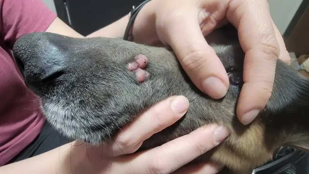
Dog histiocytomas are typically quite distinctive in their appearance, making them somewhat recognisable, though professional veterinary diagnosis is always essential. They are often described as “button tumours” due to their characteristic shape. Pet owners usually first notice these bumps during petting, grooming, or bathing sessions due to their rapid onset.
Key symptoms and characteristics of a typical histiocytoma include:
- Appearance: A raised, round, or dome-shaped lump on the skin.
- Colour: Often red or pink.
- Texture: Can have a smooth, shiny, or hairless surface.
- Size: Generally small, typically less than 2.5 cm (about an inch) wide, though they can vary. They grow rapidly in the first 1-4 weeks.
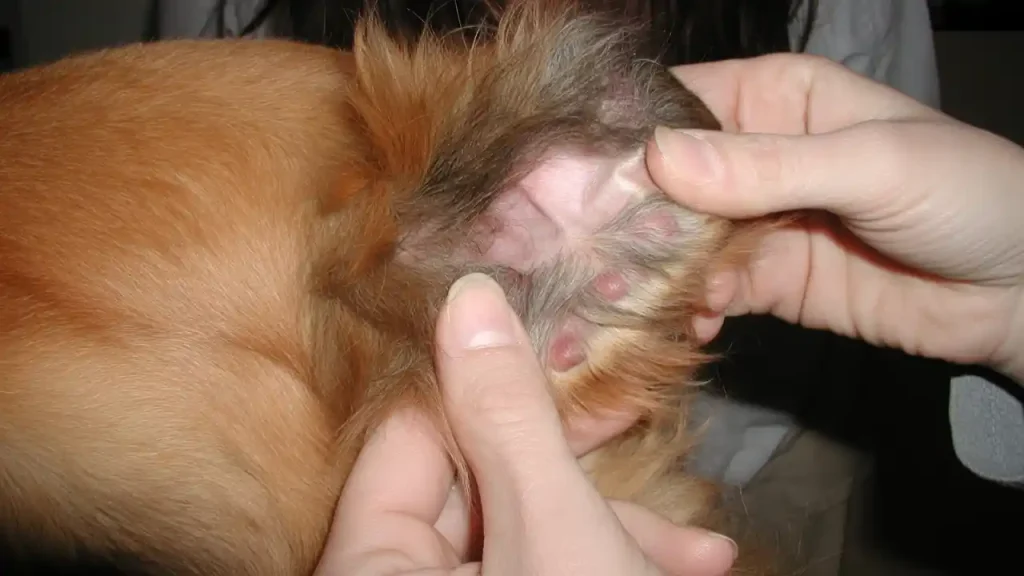
- Location: Most commonly found on the head, face, ears, and legs, but they can appear anywhere on the body.
- Solitary Nature: Usually appear as a single lump.
- Mobility: Often moves with the surrounding skin, indicating it’s not attached to underlying tissues.
While histiocytomas are generally not painful, they can become irritated, itchy, or ulcerated, especially if the dog licks, bites, or scratches the area excessively. This irritation can lead to secondary bacterial or fungal infections, which may present as an open sore with pus, bleeding, or localised swelling around the lump. If a histiocytoma is on a paw or limb, it might rub against surfaces, causing further irritation. Pet owners should monitor for any such changes, as these complications might necessitate veterinary intervention.
Diagnosing Dog Histiocytoma: From Physical Exam to Biopsy
When a pet owner discovers a new lump or bump on their dog’s skin, it’s natural to be concerned. While dog histiocytomas are often benign, it’s crucial to seek veterinary attention for any new growth to ensure an accurate diagnosis and rule out more serious conditions. Many types of skin masses can look similar, including mast cell tumours, which are malignant and require different treatment.
The diagnostic process typically begins with a thorough physical examination by your veterinarian. They will assess the size, shape, colour, location, and consistency of the mass, along with any signs of irritation, ulceration, or infection. The dog’s age and breed history will also be taken into consideration, as these are strong indicators for histiocytomas.
The most common and often sufficient diagnostic tool for histiocytomas is a fine needle aspiration (FNA) with subsequent cytological examination. This minimally invasive procedure involves using a thin needle to collect a sample of cells from the lump. The collected cells are then spread on a microscope slide and examined by a veterinarian or a veterinary pathologist. For histiocytomas, cytology typically reveals a characteristic population of round histiocytic cells. FNA is quick, relatively inexpensive, and usually causes little discomfort to the dog, often not requiring sedation.
In cases where the FNA results are inconclusive, or if the growth has an unusual appearance, is persistent, or occurs in an older dog, a biopsy may be recommended for histopathological examination. A biopsy involves removing a small piece of tissue or the entire mass (excision biopsy). This sample is then sent to a specialised laboratory where a veterinary pathologist examines the tissue architecture under a microscope. Histopathology with immunohistochemical staining provides a definitive diagnosis and can confirm that the tumour is indeed a benign histiocytoma, differentiating it from other potentially malignant skin tumours. A biopsy may require local anaesthetic or sedation, depending on the location and size of the mass.
Effective Treatment Options for Canine Histiocytoma
The good news for pet owners is that dog histiocytomas often require minimal intervention due to their benign nature and tendency for spontaneous regression. The primary approach often involves a period of watchful waiting, but other treatments are available if needed.
1. Watchful Waiting (Benign Neglect): For many histiocytomas, especially in young Dog Histiocytoma where the diagnosis has been confirmed, the most common and appropriate approach is active monitoring. The Dog Histiocytoma immune system will typically recognise and eliminate the tumour on its own within two to three months. During this time, owners must prevent the dog from licking, chewing, or scratching the mass, as this can lead to irritation, ulceration, and secondary infection. An Elizabethan collar (e-collar) may be used to prevent self-trauma.
2. Surgical Removal (Excision): Surgical excision is a highly effective treatment option, providing the fastest route to healing. It is generally recommended in several scenarios:
- If the histiocytoma does not regress on its own within two to three months (or up to 10 months).
- If the tumour is particularly large, ulcerated, infected, bleeding, or causing pain and discomfort to the Dog Histiocytoma.
- If the mass is located in an area where it interferes with daily activities or is prone to constant irritation (e.g., on an eyelid, paw, or in a joint area).
- If there’s any doubt about the diagnosis, a full excision allows for comprehensive histopathological examination of the entire mass.
Surgical removal provides immediate resolution and allows for definitive confirmation of the benign nature of the growth.
3. Cryotherapy: For smaller tumours, especially those in delicate areas where traditional surgery might be difficult (such as an eyelid), cryotherapy (freezing the lesion) can be an option. This method requires special equipment and should be discussed with your veterinarian.
4. Management of Secondary Infections: If the histiocytoma becomes ulcerated or infected due to licking or trauma, your veterinarian may prescribe topical or systemic antibiotics to clear the infection. Keeping the area clean with mild saline and preventing further self-trauma are also crucial for healing.
What to Avoid: It’s generally recommended to avoid using corticosteroids for histiocytomas, as these drugs can suppress the immune response that is necessary for the tumour’s natural regression.
The choice of treatment will depend on various factors, including the Dog Histiocytoma age, the tumour’s size and location, whether it’s causing discomfort, diagnostic confirmation, and the owner’s preference. Close collaboration with your veterinarian is essential to determine the best course of action. For further information on general pet health and care, you can consult reliable resources like Wikipedia’s page on dog health.
| Treatment Option | Description | When It’s Recommended | Pros | Cons |
|---|---|---|---|---|
| Watchful Waiting | Monitoring the growth for spontaneous regression over 2-3 months. | Most confirmed histiocytomas, especially in young Dog Histiocytoma , are not irritating or infected. | No surgery, no anaesthesia, natural resolution by the immune system. | Requires patience, risk of irritation/infection, and potential for misdiagnosis if not confirmed. |
| Surgical Removal | Complete excision of the tumour under anaesthesia. | Non-regressing tumours (>3 months), large/irritating masses, infected/ulcerated lesions, problematic locations, and diagnostic uncertainty. | Immediate resolution and a definitive diagnosis via histopathology prevent complications. | Requires anaesthesia, surgical recovery, and potential for sutures/scarring. |
| Cryotherapy | Freezing the tumour to destroy cells. | Small tumours, especially in delicate areas where surgery is difficult. | Minimally invasive procedures may avoid extensive surgery. | Requires specialised equipment, may not be suitable for larger or deep tumours, and has potential for local irritation. |
| Antibiotics | Oral or topical medication to treat bacterial infections. | When the histiocytoma becomes secondarily infected due to licking or ulceration. | Treats secondary complications and reduces discomfort. | It does not treat the histiocytoma itself and requires additional medication. |
Prognosis and Long-Term Management of Histiocytoma
The prognosis for a Dog Histiocytoma diagnosed with cutaneous histiocytoma is overwhelmingly excellent. In the vast majority of cases, these tumours are benign and either regress spontaneously or are completely cured by surgical removal. The chance of recurrence in the same location after surgical removal is very low, typically less than 1%.
Even if left untreated and allowed to regress naturally, the immune system effectively eliminates the tumour, leaving the skin as it was before the growth appeared. This process involves T-lymphocytes infiltrating and destroying the histiocytoma cells. While spontaneous regression is the norm, it’s crucial to differentiate histiocytomas from other, more aggressive histiocytic disorders or other types of skin cancers. Misdiagnosis could lead to a delay in treating a more serious condition, which could have a poor prognosis.
Long-term management primarily involves continued vigilance from pet owners. Even after a histiocytoma has resolved, it’s a good practice to regularly check your dog’s skin health for any new lumps or bumps. While it’s unusual for more than one histiocytoma to appear on the same Dog Histiocytoma or for a new one to appear later at a different site, pet owners should always report any new skin growths to their veterinarian. In very rare instances, multiple histiocytomas or unusual behaviour (like spread to local lymph nodes) could indicate a more complex condition such as cutaneous Langerhans cell histiocytoma, which is more common in Shar Peis and behaves differently from a typical histiocytoma.
Post-treatment care, whether following watchful waiting or surgical removal, focuses on ensuring the area heals properly and preventing complications. If a tumour was surgically removed, keeping the incision site clean and dry and preventing the Dog Histiocytoma from licking or scratching it are paramount. Any signs of infection, significant swelling, or bleeding should be reported to your veterinarian immediately. For naturally regressing tumours, monitoring for secondary infections or persistent irritation is key. An orthopaedic dog histiocytoma bed might help cushion areas like legs if the histiocytoma were irritating, rubbing against surfaces.
Prevention and Supporting Your Dog Histiocytoma Skin Health
Given that the exact cause of dog histiocytomas is not fully understood, and a genetic component is suspected, specific prevention strategies are difficult to define. There are no known vaccines or dietary interventions that can reliably prevent these tumours.
However, pet owners can focus on general skin health and proactive monitoring to ensure any growths are detected early and managed appropriately. Here are some recommendations for prevention and long-term care:
- Regular Skin Checks: Routinely inspect your Dog Histiocytoma skin during grooming or petting. Pay close attention to areas where histiocytomas commonly appear, such as the head, face, ears, and legs. Early detection allows for timely veterinary assessment.
- Prompt Veterinary Consultation: Any new lump or bump on your Dog Histiocytoma skin should be evaluated by a veterinarian. While it may turn out to be a benign histiocytoma, it’s crucial to rule out more serious conditions.
- Preventing Self-Trauma: If a histiocytoma develops, prevent it from licking, chewing, or scratching it. Self-trauma can lead to ulceration, infection, and increased discomfort. An Elizabethan collar (e-collar) may be necessary.
- Maintain Good Hygiene: Keeping your dog’s histiocytoma skin clean and healthy can help prevent secondary bacterial infections, especially if a lump becomes irritated or ulcerated. Mild saline solutions can be used to clean ulcerated areas.
- Balanced Diet and Overall Health: A nutritious diet and a healthy lifestyle support a robust immune system, which is crucial for the body’s ability to fight off and regress growths like histiocytomas. While not a direct preventative, overall wellness contributes to better health outcomes.
- Awareness of Breed Predispositions: If you own a breed known to be predisposed to histiocytomas (e.g., boxers and dachshunds), be extra vigilant with skin checks and discuss any concerns with your veterinarian.
While you may not be able to prevent histiocytomas from forming, proactive care and prompt veterinary attention ensure that these typically benign growths are identified correctly and managed effectively, minimising discomfort for your canine companion. The primary goal is to prevent irritation and infection while waiting for natural regression or to intervene surgically if necessary.
Conclusion
Dog histiocytoma, often affectionately known as a “button tumour”, is a common and usually benign skin growth primarily affecting young dogs. Originating from Langerhans cells in the skin, these rapidly appearing lumps typically resolve spontaneously within a few months, thanks to the Dog Histiocytoma own immune system. While their sudden onset and appearance can be concerning for pet owners, understanding their benign nature and predictable course is key to managing them effectively.
Accurate diagnosis by a veterinarian, usually through a fine needle aspiration (FNA) or biopsy, is crucial to differentiate histiocytomas from other, more serious skin tumours that may appear similar. For most cases, watchful waiting and preventing self-trauma are sufficient. However, surgical removal or cryotherapy may be recommended if the tumour is persistent, large, ulcerated, infected, or located in an area causing significant irritation.
The prognosis for dog histiocytoma with histiocytomas is excellent, with a very low chance of recurrence. By maintaining regular skin checks, seeking prompt veterinary advice for any new growths, and following recommended care, pet owners can ensure their canine companions remain healthy and comfortable. While it’s impossible to prevent every histiocytoma, informed and proactive care is the best approach to managing these common, yet usually harmless, skin conditions.