Dog Keratin Growth: 10 Essential Facts About Growths Under the Chin (2026 Guide)
Table of Contents
Dog keratin growth and various formations under a dog’s chin are common concerns that can prompt pet owners to seek veterinary attention. While many growths are benign and resolve with minimal intervention, others may signal underlying health issues, including serious conditions that require immediate medical care. Understanding the role of keratin in canine health, recognizing the signs of abnormal keratin production, and being aware of the different types of growths that can appear under a dog’s chin are crucial steps in ensuring your furry companion’s well-being. This comprehensive guide will delve into these topics, providing essential information to help owners identify potential problems and understand when professional veterinary consultation is indispensable.
Understanding Keratin and Its Vital Role in Canine Health
Keratin is a fundamental structural protein, acting as the primary building block for a dog’s skin, hair, nails, and claws. This fibrous protein is insoluble in water and organic solvents, forming a tough, protective outer layer that is critical for overall canine health. Its presence establishes the structural integrity for coat texture, nail hardness, and the durability of paw pads. The skin, being the largest organ, relies heavily on keratin to form a robust barrier, safeguarding the Dog from external threats such as chemicals, environmental stressors, trauma, and even ultraviolet light.
Beyond its protective function, keratin plays a pivotal role in preventing water loss through the skin, thereby maintaining proper hydration. The process of keratinization involves specialized skin cells called keratinocytes, which continuously produce keratin, leading to the constant shedding and replacement of old skin cells. This continuous renewal ensures the skin remains healthy and functional. When this intricate process is disrupted, either through overproduction or issues with its structural integrity, it can lead to various dermatological problems that manifest as visible changes in a dog’s coat, nails, or skin, highlighting keratin’s indispensable contribution to a dog’s overall vitality and immune system function.
When Keratin Goes Awry: Hyperkeratosis in Dogs
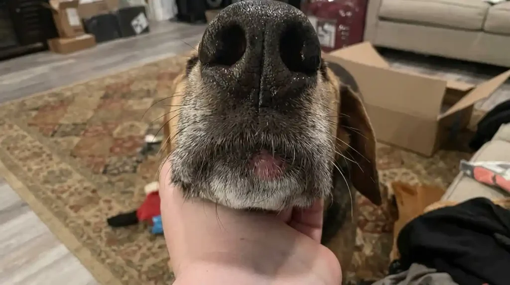
Hyperkeratosis in Dog Keratin is a common dermatological condition characterized by an excessive production and accumulation of keratin, resulting in thickened, dry, and often painful areas of skin. This overstimulation of keratin production can lead to significant changes in the texture and appearance of affected areas.
While hyperkeratosis can occur on various parts of the body, it is most frequently observed on the nose (nasal hyperkeratosis) and paw pads (paw pad hyperkeratosis), and sometimes on pressure points like the elbows. Symptoms typically include the development of rough, dry, and hardened skin, which may crack, form fissures, or develop a crusty or scaly texture. In severe cases, these cracks can bleed and become sensitive to touch, causing discomfort, pain, and even lameness, particularly if the paw pads are affected. Owners might also notice discoloration of the affected areas, hairlike projections on the paw pads, or excessive licking and chewing due to irritation.
Causes of Hyperkeratosis:
- Genetic and Hereditary Predisposition: Some dog breeds are genetically predisposed to hyperkeratosis, with the condition appearing relatively early in life, often between 4 and 9 months of age. Breeds such as English Bulldogs, French Bulldogs, Boxers, Labrador Retrievers, Cocker Spaniels, Irish Terriers, and Dogues de Bordeaux are known to be more susceptible.
- Age-Related Changes: Hyperkeratosis is more prevalent in middle-aged and senior dogs. As Dog Keratin age, their skin tends to thicken, particularly at pressure points, leading to the formation of calluses. This age-related thickening is a common cause of excess keratin production.
- Nutritional Deficiencies: A significant cause can be zinc-responsive dermatosis, a condition where a dog’s body struggles to absorb or properly utilize zinc. Since zinc is crucial for healthy skin, its deficiency disrupts the normal function of keratinocytes, prompting them to produce excess keratin. Zinc supplements are often prescribed in such cases.
- Underlying Health Conditions: Hyperkeratosis can also be a symptom of more serious underlying diseases. These include autoimmune disorders such as Pemphigus foliaceus and lupus erythematosus, hormonal imbalances like Cushing’s disease, viral infections like the canine distemper virus, or parasitic infestations such as leishmaniasis. In older Dog Keratin, hyperkeratosis of the paw pads can even be associated with chronic liver disease or pancreatic tumors, a condition known as hepatocutaneous syndrome.
While hyperkeratosis is generally not life-threatening, it can significantly impact a dog’s comfort and quality of life. The thickened, cracked skin can be painful and vulnerable to secondary bacterial or fungal infections, making prompt diagnosis and management essential. Regular monitoring and maintaining overall skin and coat health are vital for dogs prone to this condition.
Unraveling Growths Under a Dog Keratin Chin: Common Concerns
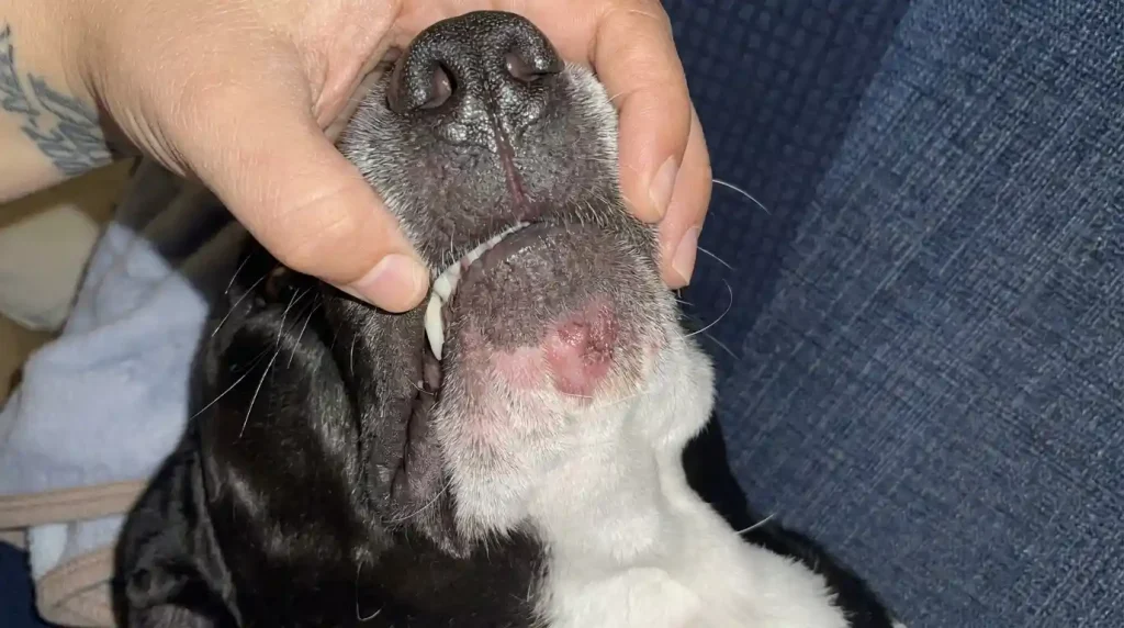
Discovering a lump or bump under your Dog Keratin chin can be a source of significant anxiety for pet owners. These growths vary widely in nature, from harmless fatty deposits to more serious conditions requiring immediate veterinary attention. While you cannot determine the severity of a growth simply by looking at it, understanding the common types can help you know what to watch for.
Benign Growths:
- Lipomas: These are non-cancerous fatty tumors that are typically soft, movable, and often feel squishy to the touch. While they can appear anywhere on the body, including under the chin, they are most common in older and overweight Dog Keratin, often found on the trunk and legs. Lipomas are generally harmless, but they can grow quite large and, depending on their location, may interfere with movement or become bothersome.
- Sebaceous Cysts/Adenomas: These growths arise from blocked oil (sebaceous) glands in the skin. They can appear as small, pimple-like bumps, often white to pink in color with irregular borders. If they rupture, they may release a thick, white, or paste-like, greasy material. While typically benign, they can become inflamed or infected. Certain breeds, such as Cocker Spaniels, are more prone to sebaceous cysts.
- Warts (Papillomas): Caused by the canine papillomavirus, warts are benign growths that often have a distinctive cauliflower-like appearance. They are more common in young Dog Keratin with developing immune systems and in older dogs with weakened immunity, frequently appearing around the mouth, muzzle, and lips. Most papillomas resolve spontaneously as the dog’s immune system clears the virus.
- Histiocytomas: These are benign skin growths that commonly manifest as red, raised, button-like lumps. Histiocytomas are most frequently observed in young Dog Keratin (typically under two years of age) and usually appear on the front half of the body, including the head. They often regress spontaneously over time without specific treatment.
- Skin Tags (Acrochordons): These are generally harmless growths resulting from an overgrowth of connective tissue in the skin. They typically appear as small flaps or stalk-like protrusions, often the same color as the surrounding skin. Skin tags are more common in older Dog Keratin and in areas where skin rubs against skin.
- Follicular Cysts: These benign, circular masses originate from a blockage of a hair follicle. They can contain a thick, often gray or yellow-brown, material. While often small, they can grow to several centimeters, become itchy or painful, and may rupture, leading to inflammation and potential infection.
Inflammatory/Infectious Growths:
- Abscesses: Abscesses are painful pockets of pus that form under the skin, usually due to a bacterial infection. Common causes include bite wounds from other animals, penetrating injuries from foreign objects (like splinters or grass seeds), or infections originating from dental issues. Abscesses typically present as swollen, warm, red, and tender lumps, and they may rupture, releasing pus and a foul odor. These require prompt veterinary treatment for drainage and antibiotics.
- Dog Keratin Acne (Folliculitis/Furunculosis): Similar to human acne, Dog Keratin acne is an inflammatory disorder that results in red, pimple-like bumps, often found on the chin, muzzle, and lips. It is more common in younger, short-haired breeds and can develop when short hairs push below the surface, becoming inflamed (folliculitis), or when hair follicles become infected (furunculosis). If left untreated, the bumps can worsen and become infected.
- Allergic Reactions/Hives: Sudden onset of small, red, itchy bumps, sometimes accompanied by swelling of the face, can indicate an allergic reaction. These can be triggered by insect bites, environmental allergens, or food sensitivities. While often benign, severe allergic reactions can be life-threatening and warrant immediate veterinary care.
Malignant (Cancerous) Growths:
Although most lumps are benign, some growths under the chin can be malignant and require aggressive treatment.
- Mast Cell Tumors: These are among the most common skin tumors in Dog Keratin and can vary significantly in their appearance and behavior. They can cause localized swelling and irritation, and their severity can range from benign to highly aggressive.
- Melanomas: Often darkly pigmented, melanomas can be aggressive and have a high rate of metastasis. While commonly found on the lips, toes, and scrotum, they can also appear as a black spot or lump under the chin or in the mouth. Rapid growth, ulceration, or bleeding are red flags.
- Squamous Cell Carcinomas (SCC): These tumors often appear as ulcerated, non-healing sores and can be highly aggressive. In the oral region, they are frequently found on the gums or tongue and can invade underlying bone.
- Fibrosarcomas: These are firm, deep-seated tumors that may invade surrounding tissues. They can be locally aggressive, though they are often slower to metastasize than some other types of cancer.
- Lymphoma: This cancer affects the lymph nodes, and swollen submandibular lymph nodes located under the chin can be a sign of lymphoma. Lymph nodes may also become enlarged due to infection or other immune responses.
Any new lump or change in an existing growth should be evaluated by a veterinarian to ensure proper diagnosis and timely intervention.
The Diagnostic Process for Canine Growths
When a Dog Keratin owner discovers a lump, bump, or area of abnormal keratin growth, a thorough veterinary evaluation is the crucial next step. The diagnostic process typically begins with a comprehensive physical examination, where the veterinarian will observe and palpate the growth, noting its size, shape, color, texture, mobility, and any associated pain or discomfort. This initial assessment provides valuable clues about the nature of the growth.
To obtain a definitive diagnosis, several diagnostic tests may be recommended:
- Fine Needle Aspirate (FNA) and Cytology: An FNA is a minimally invasive and relatively inexpensive procedure often performed during a regular outpatient visit. A small needle is used to collect a sample of cells from the mass, which are then examined under a microscope (cytology) by a veterinary pathologist. This can often distinguish between fatty tumors, cysts, inflammatory lesions, and some types of cancer.
- Biopsy and Histopathology: If an FNA is inconclusive or if the veterinarian suspects a more serious condition, a biopsy may be performed. This involves surgically removing a small piece or the entire growth for histopathological examination by a specialist. A biopsy provides a more definitive diagnosis and can determine if a growth is benign or malignant, as well as its specific type.
- Blood Work and Urinalysis: These baseline tests can help rule out underlying systemic diseases that might be contributing to skin conditions or growths, such as infections, hormonal imbalances (e.g., Cushing’s disease), or organ dysfunction (e.g., liver or kidney disease).
- Imaging (X-rays, Ultrasound, CT/MRI): For deeper-seated growths, those affecting bone, or suspected malignant tumors, imaging techniques may be employed. X-rays can assess bone involvement, ultrasound can visualize soft tissue structures, and advanced imaging like CT or MRI scans can provide detailed views to determine the extent of a tumor and check for metastasis (spread) to internal organs like the lungs.
Veterinary oncologists, such as Dr. Sue Ettinger, advocate for a proactive approach known as “See Something. Do Something: Why Wait? Aspirate”. This initiative encourages early detection and intervention for skin masses. According to these guidelines, if a skin growth has been present for a month and is approximately 1 cm in diameter (about the size of a pea), it should be aspirated or biopsied. Early diagnosis, especially when tumors are small, can significantly improve treatment outcomes and may allow for less invasive surgical procedures. Regular monthly checks by owners, coupled with prompt veterinary evaluation of any new or changing growths, are essential for optimal pet health.
| Type of Growth | Typical Appearance | Common Location(s) | Typical Diagnosis Method(s) |
|---|---|---|---|
| Hyperkeratosis | Thickened, dry, crusty, cracked skin; hairlike projections | Nose, paw pads, elbows, chin (less common) | Physical exam, breed history, rule out underlying disease (blood work, biopsy) |
| Lipoma | Soft, movable, non-painful fatty lump under skin | Trunk, legs, sometimes chin; common in older/overweight Dog Keratin | Palpation, Fine Needle Aspirate (FNA) |
| Sebaceous Cyst/Adenoma | Pimple-like bump, may rupture and release cheesy/greasy material | Anywhere, common in breeds with fine hair (e.g., Cocker Spaniels) | Physical exam, FNA |
| Wart (Papilloma) | Cauliflower-like growth, often small | Mouth, lips, muzzle, sometimes chin; common in young/old Dog Keratin | Visual inspection, biopsy if atypical |
| Abscess | Swollen, painful, warm, red lump; may ooze pus | Anywhere, often near wounds (e.g., bites |