Dog Skin Lumps: 5 Crucial Tips to Avoid Dangerous Mistakes Early
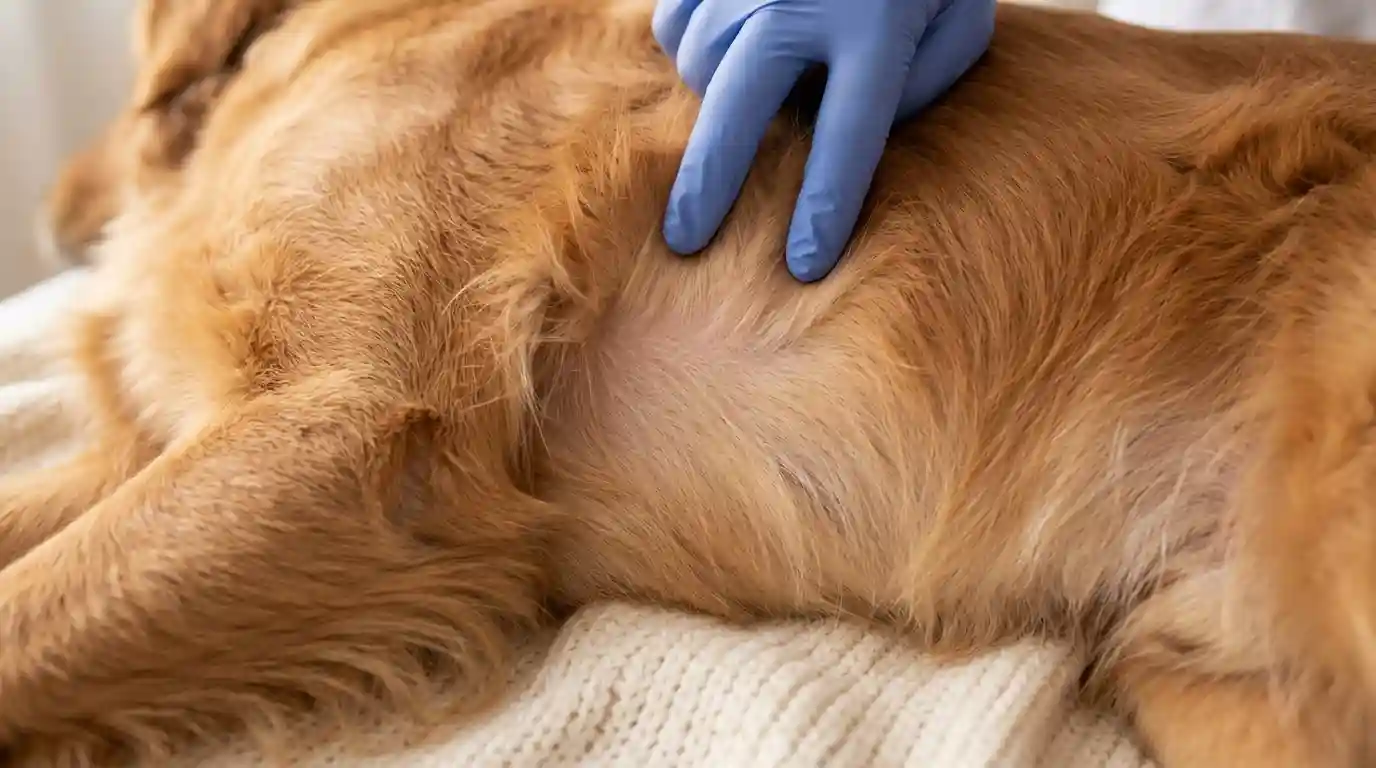
Table of Contents
Discovering a new lump, bump, or growth on your dog’s skin can be an alarming experience for any pet owner. Because our dogs’ skin is their largest organ and acts as a primary shield against the environment, it is highly prone to developing a wide variety of surface anomalies.
The good news is that the vast majority of dog skin growths are entirely benign (non-cancerous). However, because some malignant (cancerous) growths can perfectly mimic harmless tissue on the surface, understanding what these growths are, what causes them, and when to seek veterinary intervention is crucial for your dog’s long-term health.
This comprehensive, medically vetted guide breaks down the most common types of canine skin growths, their distinct characteristics, diagnostic protocols, and treatment options.
1. Demystifying the Terminology: What Exactly Is a Skin Growth?
In veterinary medicine, the terms “growth,” “lump,” “bump,” “mass,” and “tumor” are often used interchangeably by pet owners, but they carry distinct diagnostic meanings.
- Growth / Mass: General terms used to describe any abnormal swelling, cluster of cells, or prominent pocket of tissue rising above or sitting just beneath the skin surface.
- Cyst: A non-cancerous, sac-like pocket of tissue that is typically hollow and filled with fluid, air, or semi-solid material (such as keratin or sebum).
- Tumor: A solid mass of tissue formed by an abnormal collection of cells. A tumor can be either benign (localized, slow-growing, and non-invasive) or malignant (invasive, fast-growing, and capable of spreading).
- Neoplasia: The scientific medical term for the uncontrolled, abnormal growth of cells.
2. Comprehensive Classification of Common Benign Dog Skin Growths
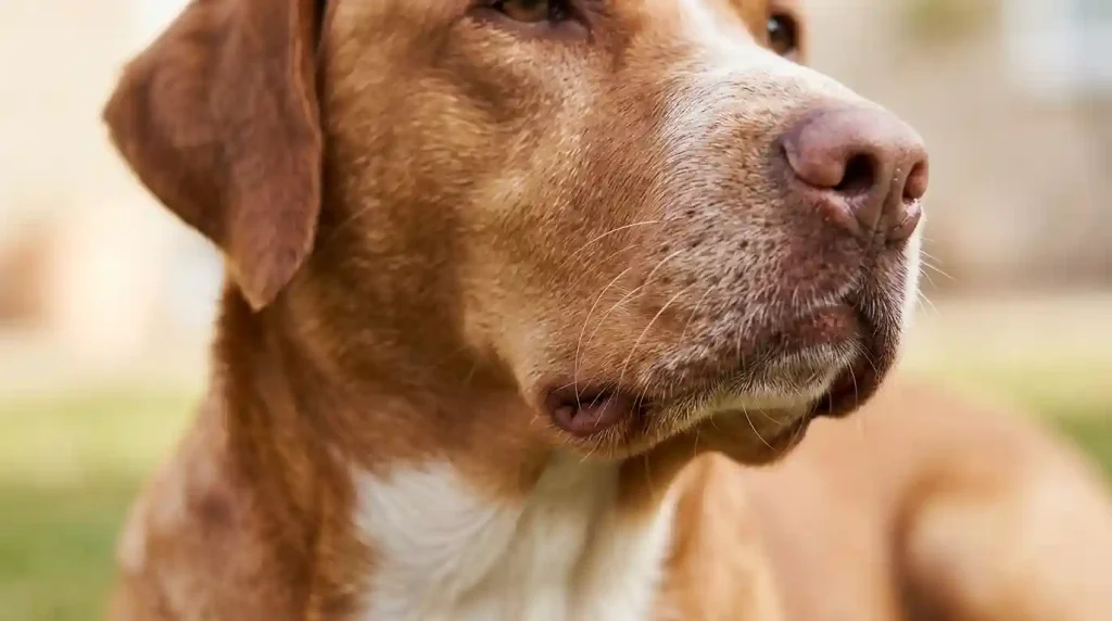
Benign growths do not invade surrounding tissues or metastasize (spread) to distant organs. While they are usually harmless, they may require management if they grow large enough to restrict movement, cause discomfort, or become repeatedly infected.
Lipomas (Fatty Tumors)
Lipomas are arguably the most common subcutaneous (under the skin) growths found in veterinary practice, particularly in middle-aged and senior dog skin.
- What they feel like: Soft, squishy, dome-shaped, and easily moveable under the skin. They are non-painful and tend to grow very slowly.
- Common locations: The chest, abdomen, armpits, and upper limbs.
- Risk factors: Older dog skin, sedentary pets, and overweight or obese dog skin are highly predisposed. Breeds like Labrador Retrievers, Golden Retrievers, and Weimaraners see them frequently.
Sebaceous Hyperplasia & Adenomas
These growths occur when the skin’s oil-producing glands (sebaceous glands) become overactive or blocked, leading to localized cellular overgrowth.
- What they look like: Small, raised, pinkish-white, or flesh-colored nodules that frequently resemble a miniature head of cauliflower or a classic wart. They can occasionally secrete a white, waxy substance if irritated.
- Common locations: Head, neck, back, and limbs.
- Risk factors: Exceptionally common in senior dog skin of all breeds, notably Cockers Spaniels, Poodles, and Schnauzers.
Canine Papillomas (Warts)
True warts are viral in nature, caused by the highly contagious canine papillomavirus.
- What they look like: Jagged, rough, flesh-colored, or white growths that sprout quickly. They are highly transmissible through direct contact or shared toys.
- Common locations: Inside and around the mouth, muzzle, lips, and occasionally around the eyes.
- Risk factors: Young puppies (under 2 years old) with developing immune systems or immunocompromised senior dog skin.
Histiocytomas (“Button Tumors”)
Histiocytomas are unique, benign masses arising from histiocytes—a specific subset of immune cells located in the skin.
- What they look like: Distinct, bright red, hairless, dome-shaped button-like lumps. They appear suddenly and can look alarming because they occasionally ulcerate or weep.
- Common locations: Face, ears, muzzle, and the extremities of the limbs.
- Risk factors: Primarily seen in young dog skin under 3 years of age. Predisposed breeds include Boxers, Bulldogs, Scottish Terriers, and Greyhounds. Note: These often self-resolve and disappear naturally within 2 to 3 months as the immune system matures.
Skin Tags (Acrochordons)
Skin tags are completely harmless, superficial overgrowths of fibrous tissue.
- What they look like: Small, fleshy pendulous growths attached to the body by a narrow, distinct stalk (peduncle).
- Common locations: Chest, armpits, hocks, or any area where the skin naturally rubs together.
Sebaceous Cysts
Unlike solid tumors, cysts are fluid or semi-solid structural pockets.
- What they feel like: Firm to semi-fluctuant nodules under the skin. If they rupture, they express a thick, cheese-like material made of trapped keratin and oils. They can become red and inflamed, and secondary bacterial infections are common if a dog’s skin scratches them.
3. Malignant Dog Skin Growths (Skin Cancer)
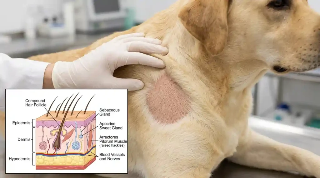
Malignant tumors are aggressive, infiltrate surrounding muscle or healthy skin tissue, and have the potential to spread through the lymphatic system or bloodstream to internal organs like the lungs and liver.
Mast Cell Tumors (MCTs)
Known universally in veterinary medicine as “The Great Imitator,” mast cell tumors are the most common malignant skin cancers in dog skin.
- Why they are dangerous: MCTs can look like absolutely anything. They can mimic a harmless lipoma, a small red bug bite, an allergic hive, or a hairless patch of skin. They can change size rapidly—expanding one day due to histamine release and shrinking the next.
- Predisposed Breeds: Boxers, Boston Terriers, Pugs, Bulldogs, Golden Retrievers, and Rhodesian Ridgebacks.
- Prognosis: Highly dependent on the pathological “grade” assigned after surgical removal. Low-grade tumors are often cured via wide surgical excision, whereas high-grade tumors require systemic chemotherapy or radiation.
Melanomas
Melanomas originate from melanocytes (pigment-producing cells).
- Cutaneous Melanoma: Melanomas found on haired areas of the skin are usually benign in dog skin. They appear as small, dark brown, or black raised nodules.
- Mucocutaneous Melanoma: Melanomas that develop in the mouth, on the lips, or within the nail beds (digital melanoma) are highly malignant, aggressively invasive, and carry a guarded prognosis, requiring prompt surgical and immunological intervention.
Squamous Cell Carcinoma (SCC)
This is a destructive skin cancer heavily linked to environmental factors.
- What it looks like: Firm, raised, irregular, and non-healing ulcerated sores or plaques that look like crusted wounds that refuse to heal.
- Risk factors: Dog skin with light-pigmented skin, white coats, or short, sparse hair coats (e.g., Dalmatians, Bull Terriers, Beagles). Prolonged sun exposure (ultraviolet radiation) on areas like the belly, nose, and eyelids is the primary driver.
Fibrosarcomas & Soft Tissue Sarcomas
These malignant tumors arise from the connective tissues beneath the skin.
- What they feel like: Deep, firm, irregular masses that feel firmly anchored or “fixed” to the underlying tissue or bone rather than sliding freely under the skin.
Summary Reference Table of Common Canine Skin Growths
| Growth Type | Primary Visual/Tactile Characteristics | Age Group Most Affected | Malignancy Risk | Common Clinical Approach |
| Lipoma | Soft, smooth, highly movable, non-painful | Middle-aged to Senior | Benign | Monitor, unless restricting movement |
| Sebaceous Adenoma | Cauliflower-like, small, pink/flesh-colored | Senior | Benign | Monitor, or remove if chronically irritated |
| Histiocytoma | Red, hairless, raised “button” appearance | Young (under 3 years) | Benign | Often self-resolves; monitor closely |
| Papilloma (Wart) | Rough, jagged, clusters around muzzle/mouth | Puppies & Seniors | Benign | Resolves via immune system or surgery |
| Mast Cell Tumor | Highly variable; can be soft, firm, red, or fluctuating | All ages | Malignant | Wide surgical excision + Histopathology |
| Squamous Cell Carcinoma | Crusted, ulcerated, raw, flat or raised sores | All ages (Sun-exposed) | Malignant | Immediate surgical removal + Margin check |
4. The Golden Rule of Veterinary Diagnosis: “See a Lump, Poke a Lump”
It is a statistical and medical impossibility to accurately diagnose a dog’s skin growth simply by looking at it or feeling it. Even the most seasoned veterinarians cannot differentiate a benign lipoma from a low-grade mast cell tumor using external palpation alone.
To find out exactly what cells compose a mass, diagnostic tests must be performed:
Fine Needle Aspiration (FNA) and Cytology
This is the standard, primary first line of defense.
- The Procedure: A tiny, sterile needle (the same size used for a standard vaccine) is inserted directly into the growth. The vet collects a microscopic pocket of cells from inside the mass, expels them onto a glass slide, stains them, and views them under a microscope.
- The Benefit: It is quick, virtually painless, minimally invasive, and can be completed during a standard consultation without sedation. It yields an immediate diagnosis for lipomas, mast cell tumors, and cysts.
Biopsy and Histopathology
If an FNA returns inconclusive results, or if the mass does not readily shed cells (poor exfoliation), a tissue biopsy is required.
- Incisional Biopsy: A small piece or core sample of the growth is surgically removed under local or general anesthesia.
- Excisional Biopsy: The entire growth is surgically excised along with a surrounding cuff of healthy tissue and sent to a veterinary pathologist.
- The Benefit: Histopathology offers a definitive diagnosis, establishes the exact structural architectural layout of the tumor, and provides a grading system to determine if the cancer has been completely removed.
5. Modern Veterinary Treatment Protocols
Once a definitive diagnosis is rendered, your veterinarian will establish an individualized treatment roadmap based on the growth’s size, location, and malignancy status.
Conservative Management (“Watchful Waiting”)
For confirmed benign growths like lipomas, skin tags, or small sebaceous hyperplasias that do not bother the dog’s skin, the standard treatment plan is simply monitoring. The mass is measured and recorded, and the pet owner tracks any changes during regular grooming routines.
Surgical Excision (Lumpectomy)
Surgery remains the gold standard treatment for most problematic or malignant masses.
- Benign Mass Surgery: The mass is removed simply for comfort or cosmetic reasons, requiring clean, narrow surgical margins.
- Malignant Mass Surgery: The veterinarian must achieve wide, deep, and clean surgical margins (often removing 2 to 3 centimeters of completely healthy tissue around and beneath the tumor). This ensures that microscopic “tentacles” of cancer cells are not left behind in the surgical bed to spark a local recurrence.
Advanced Oncological Therapies
For aggressive, high-grade skin cancers (such as advanced mast cell tumors, malignant melanomas, or soft tissue sarcomas):
- Radiation Therapy: Used locally post-surgery if the tumor was located on an extremity (like the lower leg) where wide margins were anatomically impossible to harvest.
- Chemotherapy: Systemic medications (oral or intravenous) administered to target and neutralize circulating cancer cells before they can establish secondary tumors in the internal organs.
- Immunotherapy: Advanced biological vaccines specifically engineered to stimulate the dog’s own immune system to recognize and attack specific cancer cells (highly effective in canine oral melanomas).
6. Comprehensive At-Home Tracking: How to Create a Dog Body Map
Early detection saves canine lives. As a proactive dog skin owner, the most valuable habit you can build is performing a systematic monthly “lump and bump check” combined with maintaining an accurate dog body map.
Step-by-Step Full Body Scan
Choose a relaxing time when your dog’s skin is calm. Slowly and methodically run your fingertips flat against your dog’s skin, parting the coat in thick-furred breeds.
- Muzzle and Head: Check the lips, gums, inside the ears, and around the eyelids.
- Neck and Shoulders: Feel beneath collars and harnesses, where friction can sometimes mask or irritate new skin tags.
- Chest and Abdomen: Pay careful attention to the armpits and groin areas, as well as checking all normal anatomical features (remember: both male and female dog skin have normal nipples along their abdominal wall).
- Limbs and Paws: Run your hands down each leg, checking between the toes and webbed pads.
- Tail and Perianal Area: Lift the tail to inspect the base and the sensitive skin around the anus.
Documenting with a Body Map
Draw or print a basic outline of a dog’s skin from a side and top view. When you find an anomaly, document the following metrics:
- Date: Exactly when the growth was first noticed.
- Location: Mark the exact location on your sketch.
- Dimensions: Use a rigid ruler to measure the mass in millimeters or centimeters (avoid vague comparisons like “the size of a grape,” as grapes vary wildly in size).
- Mobility: Note whether the lump slides around freely under your fingers or feels locked solidly to the body wall.
- Surface Texture: Is it soft, firm, hard, smooth, hairless, crusty, or bleeding?
Bring this map to every routine wellness exam to streamline your veterinarian’s physical assessment.
7. Red Flags: When to Seek Immediate Veterinary Attention
While you should have any new skin mass evaluated by a veterinarian at your earliest convenience, certain symptoms dictate an urgent or emergency veterinary appointment. Contact your veterinary clinic immediately if you notice a growth displaying any of the following clinical signs:
CRITICAL RED FLAGS FOR DOG SKIN GROWTHS:
1. RAPID EXPANSION: The lump doubles in size over days or weeks.
2. COLOR CHANGES: Turning angry red, deep purple, black, or mottled.
3. ULCERATION: The skin splits open, oozes pus, bleeds, or weeps.
4. ATTACHMENT: The mass feels hard and completely fixed to underlying bone/muscle.
5. DISCOMFORT: The dog’s skin constantly licks, chews, or scratches at the mass.
6. SYSTEMIC ILLNESS: New lumps accompanied by lethargy, loss of appetite,
vomiting, or localized swelling of nearby lymph nodes. vomiting,
Conclusion
Finding a growth on your dog’s skin is a call to action, not a cause for immediate panic. By understanding the common benign variants, staying vigilant with monthly full-body checks, utilizing an organized body map, and scheduling prompt diagnostics with your veterinarian, you can ensure your companion receives the highest standard of proactive care. Early detection and definitive testing remain the absolute best tools for maintaining your pet’s health, longevity, and skin comfort.