Dog Growth: 5 Types of Skin Growths Explained
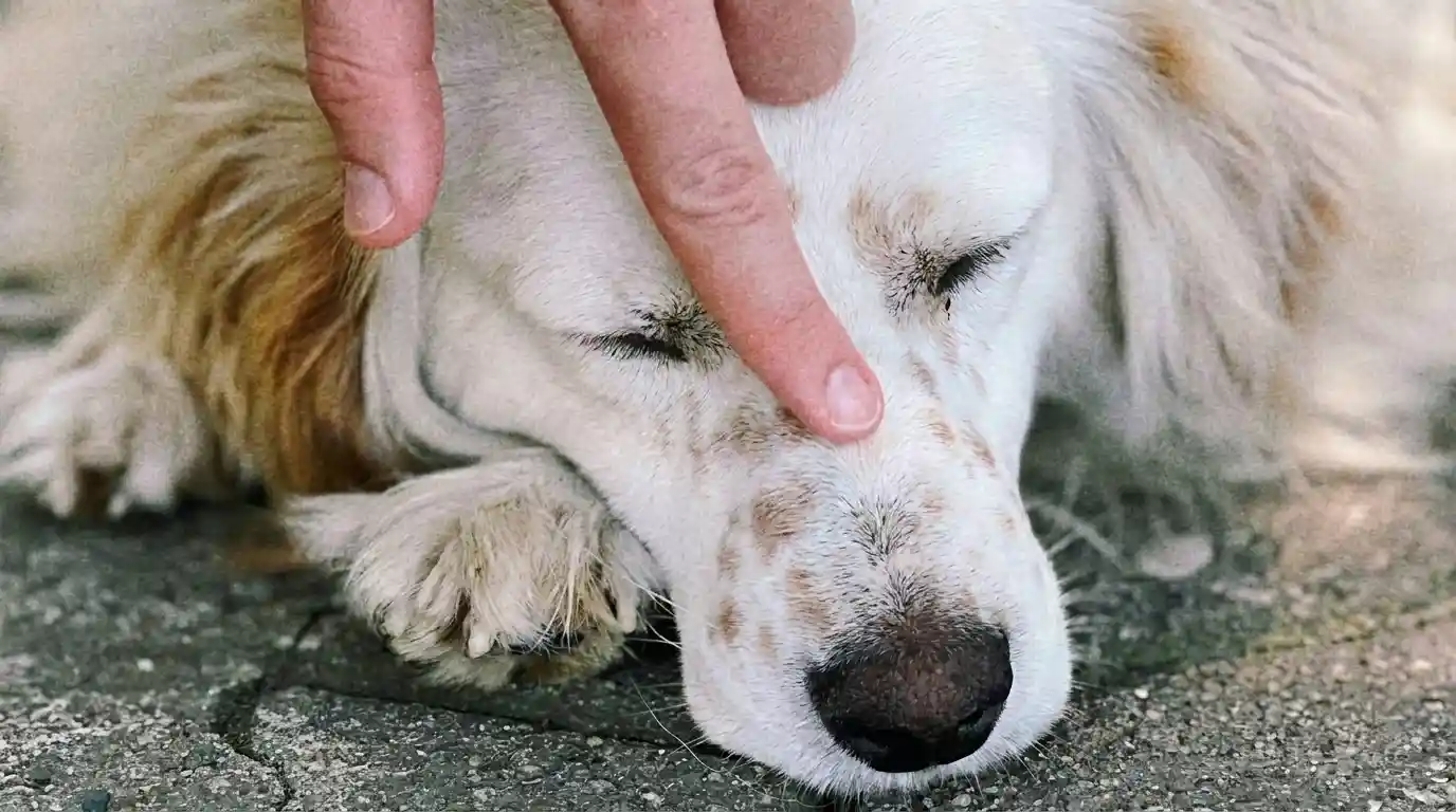
Table of Contents
Introduction to Dog Skin Growths
Dog growth on skin is a common concern for pet owners, often triggering immediate worry about their beloved companion’s health. Discovering a new lump, bump, or lesion during a routine petting session can be alarming, prompting questions about its nature and potential severity. The truth is that skin growths on dogs are extremely common and vary widely in type, ranging from completely harmless fatty deposits to those requiring prompt medical attention. The skin, being the largest organ and constantly exposed to environmental factors, is a frequent site for the development of both benign (non-cancerous) and malignant (cancerous) growths.
While many growths turn out to be benign, it is impossible to determine their nature simply by visual inspection or touch. This article aims to provide a comprehensive guide to understanding dog skin growths, covering their various types, underlying causes, diagnostic methods, and available treatment options. By understanding the common presentations and knowing when to seek veterinary evaluation, pet owners can respond appropriately and ensure the best possible outcome for their dog’s health. Early identification and intervention are consistently linked to better prognoses, especially concerning malignant lesions. Regular hands-on checks of your dog’s skin are crucial for noticing new growths promptly.
Types of Dog Skin Growths: Benign vs. Malignant
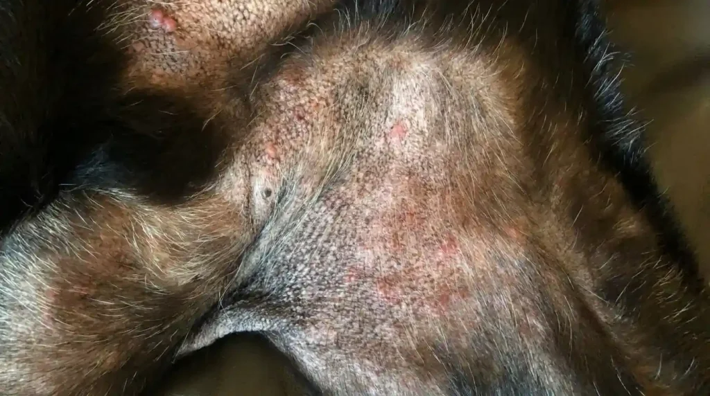
Skin growths on dogs are broadly categorized into two main types: benign and malignant. Understanding the distinction between these is critical, as it dictates the urgency and type of veterinary intervention required. Benign growths are non-cancerous and typically do not spread to other parts of the body. They may grow locally but are generally not life-threatening. In contrast, malignant growths are cancerous and have the potential to invade surrounding tissues and metastasize, or spread, to distant organs, which can be life-threatening. While a significant majority—around 60 to 80 percent—of skin tumors in dogs are benign, every new lump or bump should still be evaluated by a veterinarian to confirm its nature.
Common Benign Skin Growths
- Lipomas: These are among the most frequently encountered skin growths in dogs, composed of fatty tissue. Lipomas feel soft, movable, and are typically found just beneath the skin’s surface, often on the trunk, chest, and upper limbs. They are more common in middle-aged to older, often overweight dogs. While generally harmless, they can grow large enough to interfere with movement, necessitating surgical removal in some cases.
- Sebaceous Cysts and Adenomas: Sebaceous cysts form when oil glands become blocked, leading to fluid-filled or semi-solid sacs. They can sometimes rupture, causing local inflammation or infection. Sebaceous adenomas are benign growths of these oil-producing glands, appearing as small, sometimes greasy, white to pink masses.
- Histiocytomas: Often referred to as “button tumors,” histiocytomas are benign skin growths that commonly affect younger dogs, usually those less than two years of age. They are typically pink, raised, and can be found on the head or legs. These tumors often regress spontaneously without treatment within a few months as the dog’s immune system responds.
- Papillomas (Warts): Caused by the canine papillomavirus, these appear as small, irregular, cauliflower-like growths on the skin or around the mouth. Papillomas are more common in young dogs with developing immune systems or older dogs with weakened immunity, and many resolve on their own.
- Skin Tags (Acrochordons): These are benign fibrous growths that extend from the skin on thin stalks, usually the same color as the surrounding skin. They are generally harmless but can become irritated if caught or snagged.
- Follicular Cysts: These benign, circular masses arise from blocked hair follicles or sebaceous glands and contain a thick, often yellow-brown material. Rupture can cause pain and inflammation.
Common Malignant Skin Growths
- Mast Cell Tumors: These are the most commonly diagnosed malignant skin growths in dogs. Mast cell tumors can be highly variable in appearance, often affecting older dogs but capable of occurring at any age. They can look like small bumps, raised red patches, or ulcerated sores, and may change in size, shrinking and swelling due to histamine release. Their unpredictable nature means any new or changing lump should be promptly evaluated.
- Melanomas: Melanomas are pigmented skin tumors that can be either benign (melanocytomas) or malignant. Malignant melanomas are aggressive and often darkly pigmented, commonly developing on the lips, mouth, and nail beds, particularly in older male dogs. They grow quickly and can spread rapidly to other organs.
- Squamous Cell Carcinomas: These often appear as firm, raised, irregular, and ulcerated areas on the skin, sometimes resembling non-healing sores. They can invade surrounding tissues and are often associated with prolonged sun exposure, especially in breeds with short, white coats.
- Fibrosarcomas: These are fast-growing malignant tumors that typically appear on the trunk and legs. They can vary in appearance from lumpy under the skin to firm and fleshy deep within the muscle tissue. While they can invade local muscles, they often do not spread to distant sites.
- Angiosarcomas: Highly malignant tumors of the blood vessels that can appear as red lumps or poorly defined bruises, growing rapidly and spreading aggressively, often to the lungs and liver.
Understanding the Causes of Dog Skin Growths
Dogs develop skin growths for a multitude of reasons, reflecting the complex interaction between genetics, environment, and the aging process. The skin is continuously exposed to various factors that can trigger abnormal cell growth. Some common causes include:
- Age-Related Changes: As dogs age, they are more prone to developing skin growths. Older dogs accumulate more benign growths over time simply due to prolonged cellular activity and environmental exposure. While this doesn’t mean every new growth in a senior dog is harmless, it does indicate a natural predisposition.
- Genetic Predisposition: Breed genetics play a significant role in the likelihood of developing specific types of growths. For instance, Labrador Retrievers are particularly prone to lipomas, Boxers and Boston Terriers have higher rates of mast cell tumors, and Cocker Spaniels frequently develop sebaceous cysts. Certain breeds like Gordon Setters, Boxers, and various Terriers are at risk for hemangiomas and basal cell tumors.
- Environmental Factors: Exposure to chemicals, solar radiation, and certain viruses can contribute to the development of skin tumors. For example, squamous cell carcinomas and some angiosarcomas can be linked to sun exposure.
- Hormonal Influences: Hormonal abnormalities can also play a part in the development of some skin tumors.
- Immune Responses and Infections: Allergic reactions, insect bites, and skin infections can cause sudden bumps to form. Abscesses, for example, are painful, pus-filled swellings often caused by bacterial infections from bites or wounds. Histiocytomas, as mentioned, arise from an overgrowth of immune cells.
- Trauma: Injuries or chronic irritation can sometimes lead to localized growths or inflammatory responses that mimic tumors.
When to Consult a Veterinarian for Skin Growths
While many skin growths are benign, it is crucial for pet owners to seek veterinary evaluation for any new or changing lump, bump, or lesion on their dog’s skin. The appearance alone is not enough to determine if a growth is cancerous. Early detection and diagnosis are vital, especially for malignant tumors, as timely intervention can significantly improve prognosis and treatment outcomes.
Veterinarians recommend a proactive approach, emphasizing the “See Something, Do Something: Why Wait? Aspirate” program. This guideline suggests that if a skin growth has been present for a month and is 1 cm in diameter (about the size of a pea), it should be aspirated or biopsied.
Specific warning signs that warrant immediate veterinary attention include:
- Rapid Growth: A lump that grows quickly, doubles in size within weeks, or rapidly changes over days is a significant red flag for potential malignancy.
- Changes in Appearance: Any alteration in the size, shape, color (e.g., turning red, purple, black, or becoming multi-colored), or texture of a growth should be a cause for concern.
- Ulceration, Bleeding, or Oozing: Growths that become open sores, bleed spontaneously, or produce discharge could indicate infection, ulceration, or an aggressive tumor. Non-healing wounds are particularly worrying.
- Pain or Sensitivity: If your dog reacts with discomfort, limping, or changes in posture when a lump is touched, it might be infected, inflammatory, or a more serious underlying issue.
- Fixed or Hard Lumps: Growths that are firm and fixed to underlying tissues, rather than soft and movable, can sometimes be more concerning.
- Location: Lumps in certain critical areas like the mouth, eyelids, or near lymph nodes should be assessed promptly.
- Systemic Signs: If a growth is accompanied by other symptoms like weight loss, lethargy, loss of appetite, or difficulty moving, it suggests a systemic illness that needs urgent investigation.
Regular at-home checks are invaluable for early detection. Pet owners should routinely run their hands over their dog’s entire body, including under the legs, between the toes, around the ears, and along the tail. Documenting any new growths with measurements and photographs can provide valuable historical context for your veterinarian.
Diagnostic Procedures for Dog Skin Growths
Accurate diagnosis is paramount in determining the nature of a dog’s skin growth and formulating an appropriate treatment plan. A veterinarian cannot definitively tell if a tumor is benign or malignant simply by looking at or feeling it. Therefore, several diagnostic tools are employed to gather comprehensive information.
The diagnostic process typically begins with a thorough physical examination and a detailed clinical history. The veterinarian will assess the growth’s location, size, shape, texture, and mobility. They will also palpate nearby lymph nodes to check for enlargement, which can indicate the spread of cancerous cells.
| Diagnostic Method | Description | Key Information Provided | Advantages | Disadvantages |
|---|---|---|---|---|
| Fine Needle Aspiration (FNA) & Cytology | A small needle is inserted into the growth to collect a sample of cells. These cells are then spread on a slide and examined under a microscope by a veterinarian or a cytopathologist. | Cellular characteristics, helping to determine if a growth is benign, malignant, inflammatory, or infectious. | Rapid, minimally invasive, often done without sedation, relatively inexpensive. | May not always provide a definitive diagnosis (can be non-diagnostic), risk of missing cancerous cells if sample is too small. |
| Biopsy (Histopathology) | Surgical removal of a small piece of the mass, or the entire mass (excisional biopsy). The tissue sample is then sent to a pathology lab for detailed examination. | Definitive diagnosis, tumor type, grade, aggressiveness, and whether surgical margins are clear. Essential for confirming malignancy and guiding treatment. | Most accurate and definitive diagnostic method. | More invasive, often requires sedation or general anesthesia, results take longer, more costly. |
| Imaging Techniques | Includes X-rays, ultrasound, CT scans, and MRI. | Detailed visualization of the tumor’s size, depth, and interaction with surrounding tissues, and to check for metastasis to internal organs. | Crucial for surgical planning and staging of malignant tumors. Detects masses not visible externally. | More expensive, may require sedation. |
| Blood Tests | Routine blood work, though not directly diagnostic for skin growths, can provide information about overall health and potential systemic involvement. | General health status, organ function, and markers that might suggest inflammation or certain types of cancer. | Can highlight underlying issues or provide a baseline for treatment. | Not a definitive diagnostic tool for specific skin growths. |
Treatment Options for Canine Skin Growths
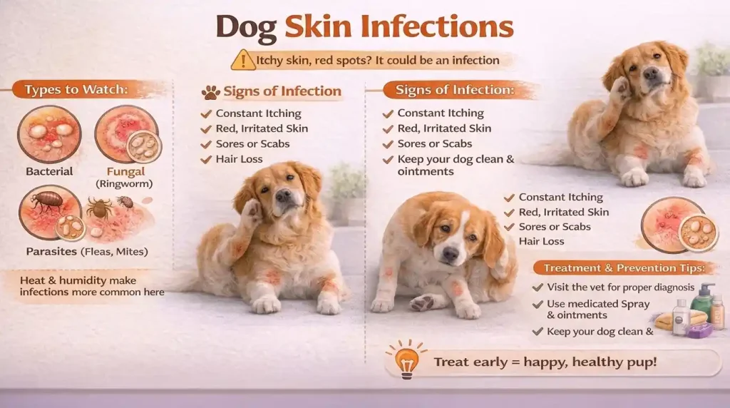
The treatment approach for a dog’s skin growth depends heavily on its type (benign or malignant), size, location, the dog’s overall health, and the owner’s preferences. A veterinarian will tailor a treatment plan based on the diagnostic findings.
Surgical Removal
Surgery is often the most common and effective treatment for both benign and malignant skin growths. For benign growths that are bothersome, growing large, or located in areas that interfere with movement, surgical excision is usually curative. The goal is complete removal with clean margins to minimize recurrence.
For malignant growths, surgery is even more critical. Veterinarians will typically remove the tumor with wider margins of surrounding healthy tissue to ensure that all cancerous cells are excised, reducing the chance of local recurrence. In some cases, partial removal may be performed to prolong the dog’s life if complete removal is not feasible. After surgical removal, the tissue is always sent for histopathology to confirm the diagnosis, determine the tumor grade, and assess the completeness of excision, which guides further treatment decisions.
Non-Surgical Treatments
For certain types of growths or in situations where surgery is not an option (e.g., due to advanced age, other health conditions, or location of the tumor), non-surgical alternatives may be considered.
- Monitoring: For small, benign, non-bothersome growths like many lipomas, skin tags, or histiocytomas that are expected to regress, a “wait and see” approach with careful monitoring may be recommended. Regular measurements and photographs are essential to track any changes.
- Cryotherapy and Laser Treatments: These methods involve freezing or using laser energy to destroy abnormal cells. They are often used for smaller, superficial benign growths like warts (papillomas) or some skin tags.
- Chemotherapy: This involves using drugs to kill cancer cells and can be administered systemically or locally. Chemotherapy is often used in conjunction with surgery for aggressive malignant tumors, or as a standalone treatment when surgery is not possible or metastasis has occurred.
- Radiation Therapy: Radiation uses high-energy rays to shrink tumors and kill cancer cells. It is typically employed for malignant tumors, either before surgery to shrink a large tumor, after surgery to eliminate any remaining cancer cells, or as a palliative measure.
- Immunotherapy: This treatment aims to stimulate the dog’s immune system to fight cancer cells. A melanoma vaccine, for instance, is available and has shown efficacy in shrinking tumor size and prolonging life expectancy for dogs with malignant melanoma.
- Intralesional Injections: For certain tumors, especially non-metastatic mast cell tumors, an FDA-approved injection (e.g., tigilanol tiglate/Stelfonta) can be directly administered into the tumor to destroy it. Experimental injections for benign tumors, such as calcium chloride solutions for lipomas, are also being explored.
It’s important to consult with a veterinary oncologist for staging and treatment recommendations if a malignancy is confirmed. For further information on veterinary oncology and cancer treatment options, pet owners can refer to resources such as the Veterinary Cancer Society website.
Prevention and At-Home Monitoring
While not all skin growths are preventable, certain practices can help minimize risks and ensure early detection, which is crucial for successful outcomes. A proactive approach to your dog’s skin health is a key component of responsible pet ownership.
- Regular At-Home Examinations: Incorporate monthly full-body checks into your routine. Gently run your hands over your dog’s entire body, paying close attention to areas that are often overlooked, such as between the toes, under the legs, around the ears, and beneath the tail. Look and feel for any new lumps, bumps, changes in skin texture, discoloration, or sores that don’t heal.
- Document Changes: If you find a new growth, note its exact location, size (measure it with a ruler if possible), shape, color, and texture. Taking clear photographs from various angles can be immensely helpful for your veterinarian to track changes over time. Keep a journal to record these observations.
- Sun Protection: For dogs with thin or light-colored fur, especially in sunny climates, limiting prolonged sun exposure can help reduce the risk of sun-induced skin cancers like squamous cell carcinoma.
- Healthy Diet and Lifestyle: A balanced diet and maintaining a healthy weight can contribute to overall immune system health and potentially reduce the incidence of certain growths, such as lipomas in overweight dogs.
- Prompt Treatment of Skin Issues: Address any skin irritations, infections, or wounds promptly to prevent secondary complications that could lead to abnormal growths.
- Regular Veterinary Check-ups: Schedule routine wellness exams with your veterinarian. These check-ups provide an opportunity for professional skin evaluations and allow your vet to identify any suspicious growths you might have missed. Discuss any concerns you have, no matter how small they seem.
Early detection is the cornerstone of effective management for dog skin growths. By being vigilant and working closely with your veterinarian, you can significantly improve the chances of a positive outcome for your pet.
Conclusion
Dog growth on skin is a prevalent issue that can range from a harmless anomaly to a serious health concern. While the discovery of any new lump or bump can be unsettling, it’s crucial for pet owners to remember that not all growths are cancerous. The diversity of skin growths in dogs necessitates a thorough and professional veterinary evaluation for accurate diagnosis and appropriate treatment. Conditions such as lipomas, sebaceous cysts, histiocytomas, and skin tags are commonly benign, often requiring only monitoring or simple removal if problematic. Conversely, malignant growths like mast cell tumors, melanomas, and squamous cell carcinomas demand prompt and aggressive intervention.
Understanding the common causes, recognizing the warning signs that warrant immediate veterinary attention, and engaging in consistent at-home monitoring are vital steps for every dog owner. Diagnostic tools like fine needle aspiration, biopsy, and advanced imaging provide veterinarians with the necessary information to distinguish between benign and malignant conditions. Treatment options vary from watchful waiting and surgical excision to more advanced therapies like chemotherapy, radiation, and immunotherapy, all tailored to the individual dog and the specific growth. By maintaining regular veterinary check-ups and actively participating in your dog’s health monitoring, you provide the best possible chance for early detection and successful management of any skin growth, ensuring a healthier and happier life for your canine companion.