Dog Keratin Growth: 10 Common Causes of Growth Under the Chin
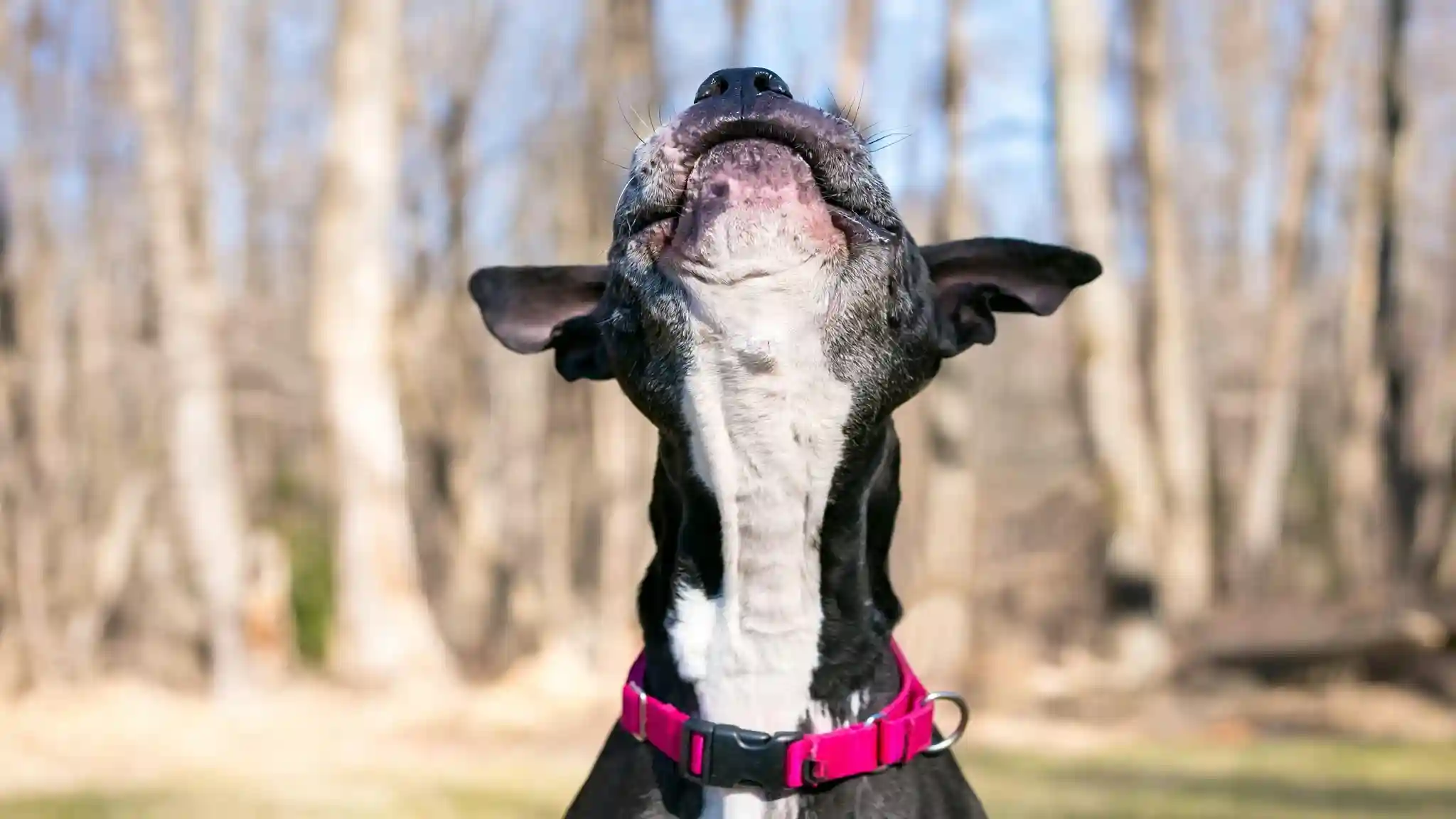
Dog keratin growth is a fundamental aspect of canine health, as keratin is the primary structural protein making up a dog’s skin, hair, and nails. This robust protein acts as a vital protective barrier, shielding the inner layers of the skin from environmental elements, preventing water loss, and guarding against injury and pathogens like bacteria, viruses, fungi, and parasites. Normal keratinization involves a continuous process where new skin cells, called keratinocytes, are produced at the base of the epidermis, migrate upwards, and progressively fill with keratin before being shed as dead cells from the skin’s surface. However, abnormalities in this process can lead to various skin conditions, often characterized by either insufficient or excessive keratin production.
Dog Keratin Growth: Understanding Healthy Skin
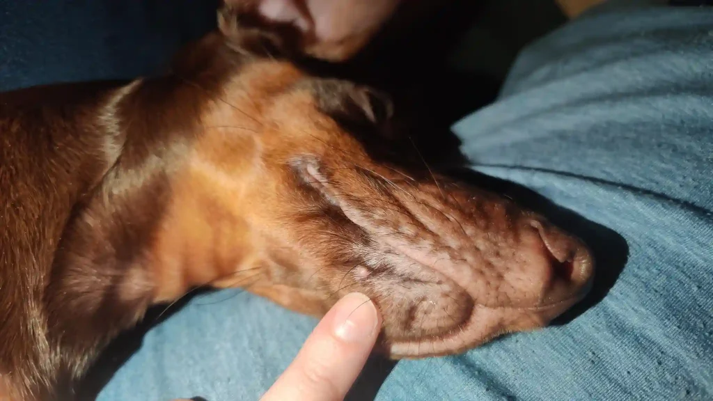
Hyperkeratosis in dogs refers to a common skin condition where there is an excessive production and accumulation of keratinocytes and keratin in certain areas of the body. This overproduction leads to thickened, dry, and sometimes rough or crusty patches of skin. While it can occur in various locations, it is most frequently observed on the nose (nasal hyperkeratosis) and paw pads (digital hyperkeratosis), and sometimes on the ears or pressure points like the elbows.
The thickened skin associated with hyperkeratosis can range from a purely cosmetic issue to a painful problem, especially if the hardened skin cracks or develops fissures. These cracks can make the Dog Keratin Growth vulnerable to secondary bacterial or yeast infections, leading to inflammation, discharge, and further discomfort. In severe cases, Dog Keratin Growth may experience pain, sensitivity in the affected area, limping, or reduced physical activity. The condition is characterized by an overabundance of keratin, a protein insoluble in water and organic solvents, which forms the outer protective layer of the skin.
Dog Keratin Growth: Hyperkeratosis and Excessive Keratin Production
The excessive production of keratin in dogs can stem from a variety of factors, including age, genetics, nutritional deficiencies, and underlying diseases. Understanding these causes is crucial for effective diagnosis and management.
Age: Hyperkeratosis is more prevalent in middle-aged and older dogs. As dogs age, their skin naturally tends to thicken, particularly at pressure points, making them more susceptible to callus formation and hyperkeratosis. Senior Dog Keratin Growth , who may spend more time lying down, can develop calluses on areas with thin skin, such as their elbows.
Breed/Genetics: Genetic predisposition plays a significant role in many cases of hyperkeratosis. Certain breeds are known to be more prone to the condition regardless of age. These include, but are not limited to, Cocker Spaniels, Dogues de Bordeaux, Irish Terriers, Labradors, English Bulldogs, French Bulldogs, Boxers, Siberian Huskies, and Alaskan Malamutes. Familial nasal and footpad hyperkeratosis are examples of hereditary keratinization disorders.
Nutritional Deficiencies: Deficiencies in certain nutrients can contribute to keratin overproduction. Zinc deficiency, for instance, can lead to abnormal keratin production and conditions like zinc-responsive dermatosis, which can manifest as hyperkeratosis. Similarly, Vitamin A responsive dermatosis is another nutritional keratinization disorder.
- Underlying Diseases: Hyperkeratosis can often be a symptom of a more significant underlying health issue. These can include:
Infectious Diseases: Conditions such as canine distemper virus and papillomavirus infection can trigger hyperkeratosis. Leishmaniasis, a parasitic condition caused by sandfly bites, can also lead to keratin hyperproduction.
Autoimmune Diseases: Autoimmune conditions like pemphigus foliaceus cause the immune system to attack skin cells, resulting in cracked and dry skin, and sometimes hyperkeratosis. Lupus erythematosus is another autoimmune condition that can cause similar symptoms.
Hormonal Imbalances: Hormonal disorders such as Cushing’s disease (hyperadrenocorticism) and hypothyroidism can also manifest with symptoms like skin hardening and thickening.
Other Keratinization Disorders: Hyperkeratosis is a specific manifestation of a broader category known as keratinization disorders, which are characterized by abnormalities in the process of epidermopoiesis, keratinization, and cell desquamation. These can be primary (hereditary, idiopathic, nutritional) or secondary (due to parasites, allergies, infections, endocrine issues, or neoplasia).
Chronic Pressure/Trauma: Constant pressure on thin skin, such as over the elbows, can lead to excessive keratin formation as a protective response.
Dog Keratin Growth: Common Causes of Hyperkeratosis
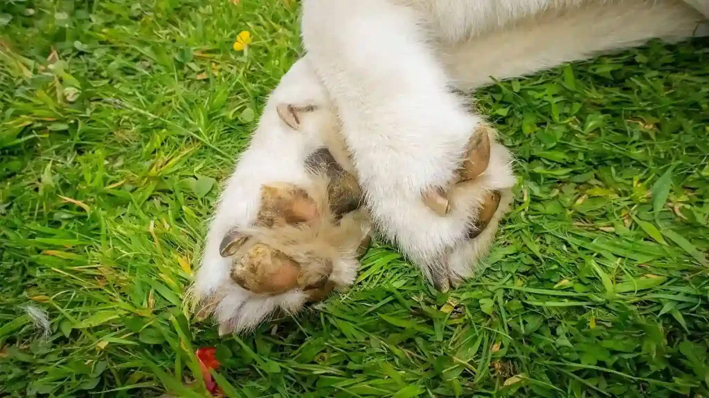
Recognizing the symptoms of hyperkeratosis early is vital for appropriate intervention and to ensure a Dog Keratin Growth comfort. The symptoms can vary in severity, from mild cosmetic changes to painful, debilitating conditions.
Common Symptoms of Hyperkeratosis:
Thickened, Dry, and Hard Skin: The most noticeable symptom is the presence of unusually thick, dry, rough, and hardened skin, typically on the paw pads and nose. This can sometimes appear as calluses.
Cracks, Fissures, and Bleeding: In more severe cases, the thickened skin can crack or develop fissures, which may bleed. These open wounds increase the risk of secondary infections.
“Hairy” or Feathered Projections: On paw pads, hyperkeratosis can sometimes manifest as abnormal tissue projections, often described as “keratin fronds” or “feathers,” giving the paw pads a rough, irregular, or “hairy” appearance.
Pain and Discomfort: The hardened, cracked skin can be sensitive to touch and cause significant pain, leading to lameness, limping, reluctance to move, or excessive licking of the affected paws.
Discoloration: Affected areas, particularly the nose, may show discoloration or loss of pigmentation, appearing lighter or even pink.
Secondary Infections: Cracks and fissures provide an entry point for bacteria and yeast, leading to secondary infections, which can cause inflammation, discharge, and a foul odor.
Diagnosis:
Diagnosing hyperkeratosis typically begins with a thorough physical examination by a veterinarian. The vet will assess the affected areas, noting the texture, appearance, and extent of the skin changes.
To determine the underlying cause and rule out other conditions, the veterinarian may recommend additional diagnostic tests:
Skin Biopsy: A skin biopsy is often the most effective way to diagnose hereditary and nutritional keratinization disorders and differentiate between various causes.
Bloodwork: Baseline blood tests may be conducted to check for underlying systemic diseases, hormonal imbalances (like Cushing’s or hypothyroidism), or nutritional deficiencies (such as zinc deficiency) that could be contributing to the hyperkeratosis.
Skin Scrapings/Cytology: To rule out parasitic infestations (e.g., Demodex mites, Cheyletiella, Sarcoptes) or secondary bacterial/yeast infections, the vet may take skin scrapings or perform cytology.
Infectious Disease Testing: Tests for infectious agents like canine distemper virus or papillomavirus may be necessary if suspected.
Allergy Testing: If allergies are suspected to be an underlying factor, allergy testing might be considered.
A precise diagnosis of the underlying cause is paramount because treatment strategies are tailored to the specific etiology.
Dog Keratin Growth Linked to Nutritional Deficiencies
Finding a lump or bump under a Dog Keratin Growth chin can be a cause for concern for many pet owners. While some growths are benign and harmless, others may indicate more serious underlying conditions, including infections, inflammatory disorders, or even tumors. It’s crucial for pet owners to have any new or changing lump evaluated by a veterinarian to receive an accurate diagnosis and appropriate treatment.
The chin area is susceptible to various types of growths due to its anatomy, frequent contact with surfaces, and the presence of hair follicles and glands. These growths can vary in size, shape, firmness, and whether they are painful or mobile.
Some of the conditions that can lead to growths under the chin are localized, while others may be manifestations of systemic issues. Differentiating between these requires a veterinary examination, as many lumps can look similar on the surface but have vastly different implications.
Dog Keratin Growth Associated with Underlying Diseases
Growths under a Dog Keratin Growth chin can be attributed to several common conditions. Here’s an overview of the most frequently encountered types:
| Type of Growth | Description | Key Characteristics / Causes | Prognosis |
|---|---|---|---|
| Canine Acne | Inflammatory disorder presenting as red bumps, pustules (pimples), or blackheads on the chin and lips. | Common in young, short-haired breeds (6 months to 1 year old). Often triggered by trauma to the chin/muzzle area, clogging hair follicles, or underlying allergies. Can lead to swelling, bleeding, or scabs. | Usually treatable with topical or oral medications; severe cases may cause scarring. Good prognosis with proper management. |
| Follicular Cysts | Masses that form within hair follicles, containing fluid or thick, cheesy material (keratin). | Develop when hair follicles become blocked due to inflammation, infection, scar tissue, or debris. Can be firm, raised, smooth, and appear on or under the skin. May rupture and become infected. | Benign (non-cancerous). Often require surgical removal if large, painful, or frequently infected. Tend not to recur if fully removed. |
| Sebaceous Cysts | Fluid-filled sacs resulting from blocked sebaceous (oil) glands that produce sebum. | Appear as smooth, round, often white or bluish bumps. Can rupture and release a thick, greasy, cottage-cheese-like, or blackish discharge. Common on the head, neck, and body. | Benign and usually harmless. May resolve on their own, but can become infected, requiring cleaning or surgical removal. Do not squeeze them at home. |
| Abscesses | Painful swellings filled with pus under the skin. | Often caused by bites, scratches, foreign objects, or infected glands/sores. Can develop rapidly and are typically painful to the touch. | Requires veterinary treatment for drainage and antibiotics to prevent infection spread. Good prognosis if treated promptly. |
| Enlarged Lymph Nodes | Swelling of the lymph nodes, which are part of the immune system. | Lymph nodes under the jaw (submandibular lymph nodes) can become enlarged due to infection, inflammation, or cancer. May feel firm and mobile. | Requires veterinary investigation to determine the underlying cause, which can range from benign infection to serious conditions like lymphoma. |
| Histiocytomas | Benign skin growths arising from immune system cells. | Usually round and red, often referred to as “button tumors”. More common in young adultDog Keratin Growth. | Typically resolve on their own within a few weeks to months without treatment. |
| Lipomas | Soft, benign fatty tumors found under the skin. | Common in middle-aged to older, often overweight Dog Keratin Growth. Usually slow-growing, squishy, and movable. | Generally harmless and do not become cancerous. Surgical removal is only recommended if they become very large or interfere with movement. |
| Salivary Cysts (Ranulas/Mucoceles) | Swelling caused by saliva leaking into surrounding tissues, forming a large, firm pocket. | Typically appear under the jaw or tongue, often feeling like a thick-walled balloon filled with fluid. Can occur in any breed and age. | Requires veterinary diagnosis, often through fine needle aspirate. Treatment may involve surgical drainage or removal of affected salivary gland. |
| Tumors (Benign and Malignant) | Abnormal cell growths, which can be non-cancerous (benign) or cancerous (malignant). | Can appear as various types of lumps. Malignant tumors may grow rapidly, change in appearance, bleed, or ulcerate. Examples include mast cell tumors, fibrosarcomas, squamous cell carcinomas. | All suspicious lumps should be checked by a vet. Early diagnosis and treatment are crucial for malignant tumors. Prognosis varies widely depending on the type and stage of cancer. |
Dog Keratin Growth and Growth Under the Chin
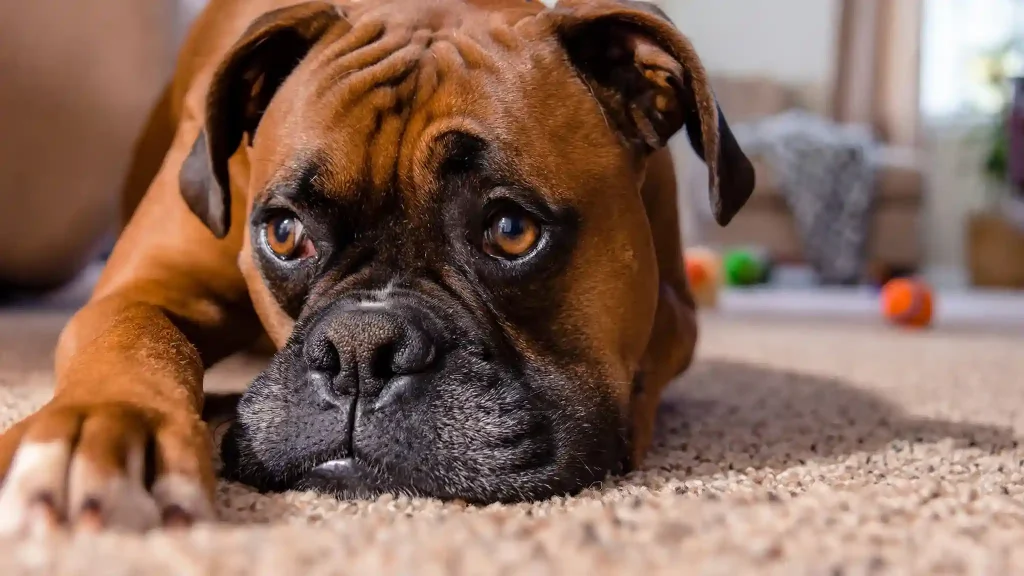
When a growth is discovered under a Dog Keratin Growth chin, the immediate step should be a visit to the veterinarian. It is impossible to accurately determine the nature of a lump simply by looking at or feeling it. A professional diagnosis is essential for determining the appropriate course of action.
Veterinary Diagnostic Process:
Physical Examination: The veterinarian will conduct a thorough physical examination, palpating the lump to assess its size, shape, firmness, mobility, and any associated pain or discomfort. They will also check for other lumps or enlarged lymph nodes elsewhere on the body.
Fine Needle Aspirate (FNA): This is a common and minimally invasive diagnostic tool. A tiny needle is inserted into the lump to collect a sample of cells, which are then examined under a microscope (cytology). FNA can often distinguish between fatty tumors, cysts, abscesses, and some types of cancerous cells, like those from a mast cell tumor or enlarged lymph node.
Biopsy: If the FNA results are inconclusive, or if cancer is suspected, a surgical biopsy may be recommended. This involves removing a larger tissue sample or the entire mass for histopathological examination by a veterinary pathologist. A biopsy provides a definitive diagnosis and is crucial for identifying the exact type of growth and whether it is benign or malignant.
Blood Tests: Blood work may be performed to assess the Dog Keratin Growthoverall health and rule out systemic diseases that could be contributing to the growth, such as infections or certain metabolic conditions.
Other Imaging: In some cases, imaging techniques like X-rays or ultrasound may be used to assess the extent of the growth, especially if it’s deep-seated or if the veterinarian suspects it might be connected to underlying structures.
Treatment Options:
Treatment for growths under the chin varies significantly depending on the diagnosis:
Observation: For benign, harmless growths like small lipomas or some histiocytomas, the veterinarian might recommend a “wait and see” approach, monitoring the lump for any changes in size, shape, or texture. Many histiocytomas, for example, resolve on their own within a few months.
- Medical Management:
Canine Acne: Mild cases can be treated with topical medications such as pet-safe shampoos or gels containing benzoyl peroxide to cleanse the skin and reduce inflammation. Topical or oral antibiotics may be prescribed for bacterial infections.
Abscesses: Require veterinary intervention for safe drainage of pus and a course of antibiotics to clear the infection.
Cysts (Follicular and Sebaceous): If uninfected and not causing discomfort, small cysts might be left alone. However, if they become inflamed, infected, or rupture, antibiotics may be needed. Larger or recurrent cysts are often surgically removed, with care taken to remove the entire cyst lining to prevent recurrence. Pet owners should never attempt to squeeze or pop cysts at home, as this can lead to rupture and secondary infection.
- Surgical Removal:
Surgical excision is a common treatment for various growths, especially those that are causing pain, interfering with function, growing rapidly, or are diagnosed as malignant.
For benign tumors like large lipomas, surgery might be considered if they hinder mobility or become problematic.
Malignant tumors (cancers) often necessitate surgical removal, sometimes followed by additional treatments like chemotherapy or radiation, depending on the type and stage of cancer.
Addressing Underlying Causes: If the growth is secondary to an underlying condition (e.g., allergies, hormonal imbalance), treating the primary cause is crucial for resolving or managing the growth. This might involve dietary changes for food allergies, environmental modifications for environmental allergies, or specific medications for endocrine disorders.
Regular cleaning of the chin and muzzle area with a gentle, pet-safe cleanser can help prevent the buildup of dirt, oil, and bacteria that contribute to conditions like canine acne. Using ceramic or stainless steel bowls instead of plastic can also reduce irritation and bacterial accumulation, thereby helping to prevent acne flare-ups. Furthermore, preventing trauma to the chin area from rough play or rubbing can reduce the likelihood of certain growths.
It is important to maintain open communication with a veterinarian about any new lumps or bumps, as early detection and appropriate management significantly improve outcomes for canine health. For further reading on various lumps and bumps in dogs, Wikipedia provides a detailed overview of dog skin diseases.
Dog Keratin Growth: Final Thoughts
The health of a Dog Keratin Growth skin, underpinned by the structural protein keratin, is a key indicator of its overall well-being. While normal keratin growth is essential for protection, excessive keratinization, or hyperkeratosis, can lead to discomfort and complications, particularly on the paw pads and nose. This condition often signals underlying issues, ranging from genetic predispositions and aging to nutritional deficiencies and serious diseases. Similarly, growths under a dog’s chin, though diverse in nature from benign cysts and acne to more serious tumors or enlarged lymph nodes, demand prompt veterinary attention. Early and accurate diagnosis through physical examination, fine needle aspirates, and biopsies is crucial for tailoring effective treatments. By understanding the causes, recognizing the symptoms, and seeking timely professional veterinary care, pet owners can ensure their canine companions receive the best possible management for both keratin-related skin issues and suspicious growths, ultimately promoting their comfort and longevity.