Dog Skin Growth: 10 Best Ways to Spot and Treat Skin Lumps
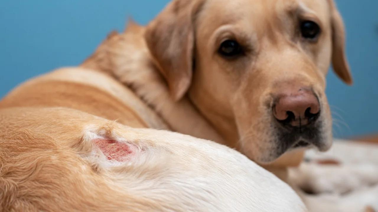
Table of Contents
Dog skin growth pictures are often the first thing concerned pet owners search for when they discover an unusual lump or bump on their beloved canine companion. Discovering a new growth on your dog can be a frightening experience, leading to immediate worry about their health. While not all skin growths are indicative of a serious underlying condition, it is crucial for pet owners to understand the various types of growths, recognize potential warning signs, and know when professional veterinary attention is necessary. This comprehensive guide aims to demystify dog skin growths, providing detailed information to help owners better understand what they might be seeing and how to respond appropriately.
Understanding the Types of Dog Skin Growths
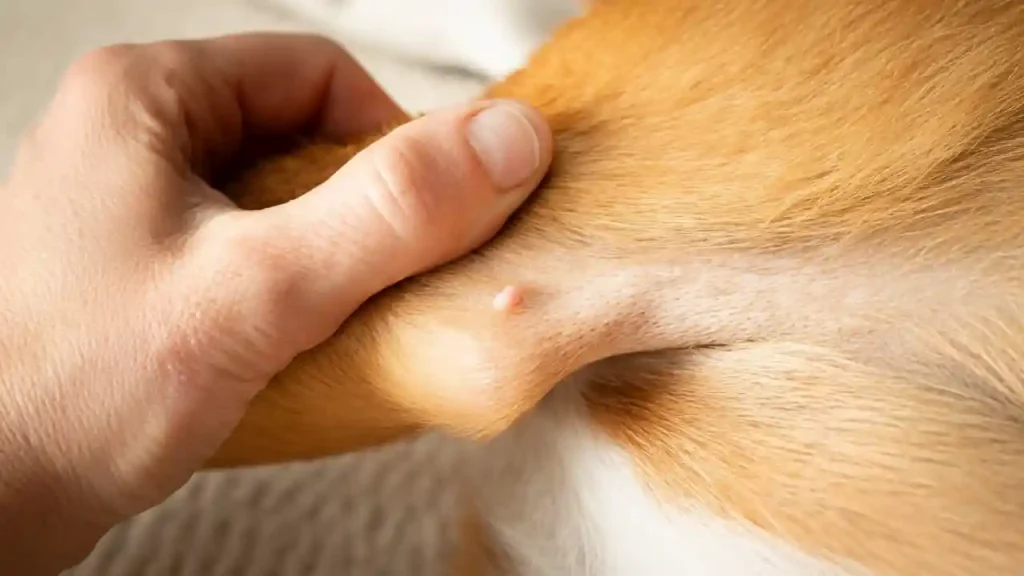
Canine skin can develop a wide array of growths, ranging from harmless benign lesions to more aggressive malignant tumors. These growths can appear anywhere on the body, vary significantly in size, texture, and color, and may develop slowly or emerge quite rapidly. The diversity of these growths makes visual identification alone challenging, underscoring the importance of veterinary examination for an accurate diagnosis. Understanding the basic categories—benign and malignant—is the first step in comprehending the potential implications of a newly discovered lump. Benign growths are non-cancerous and generally do not spread to other parts of the body, though they can sometimes cause discomfort if they grow large or are located in an area prone to irritation. Malignant growths, on the other hand, are cancerous and have the potential to invade surrounding tissues or metastasize (spread) to distant organs, posing a more serious threat to a dog’s health.
Benign Growths: What to Look For
Many skin growths found on dogs are benign, meaning they are non-cancerous and typically do not pose a life-threatening risk. However, even benign growths can cause irritation, itching, or become infected, requiring intervention. Here are some of the most common benign skin growths you might encounter on your dog:
- Lipomas: These are very common, soft, fatty tumors that typically feel movable under the skin. They are generally harmless and grow slowly, often found in older or overweight dogs. They are usually painless unless they grow very large or press on nerves.
- Sebaceous Cysts: These occur when a sebaceous gland (which produces oil for the skin) becomes blocked, leading to a build-up of sebum. They appear as firm, sometimes fluid-filled, nodules that can rupture and become infected.
- Skin Tags: Small, flesh-colored or pigmented growths that project from the skin. They are often found in areas of friction, such as the armpits or groin. They are harmless but can occasionally become irritated.
- Histiocytomas: Most often seen in younger dogs (under two years old), these are benign tumors that grow rapidly and often appear as small, firm, dome-shaped red lumps. They usually regress and disappear on their own within a few months.
- Warts (Papillomas): Caused by the canine papillomavirus, these are typically rough, cauliflower-like growths that can appear individually or in clusters, especially around the mouth or eyelids of younger dogs. They often resolve spontaneously.
- Follicular Cysts: Similar to sebaceous cysts, these arise from hair follicles and can also become blocked, leading to a firm bump that may rupture.
- Fibromas: Benign fibrous tumors that appear as firm, non-painful nodules.
While these growths are benign, it is important not to self-diagnose. A veterinarian can accurately identify these conditions through examination and sometimes additional diagnostics.
Malignant Growths: Signs of Concern
Malignant skin growths, or skin cancers, are a serious concern and require prompt veterinary attention for the best prognosis. Early detection and treatment are critical. Here are some types of malignant skin growths and general warning signs:
- Mast Cell Tumors: These are one of the most common types of skin cancer in dogs. They can appear in various forms, from a small, red, itchy lump to a larger, subcutaneous mass. They are often unpredictable in their behavior and can be aggressive.
- Melanoma: While often pigmented (dark), melanomas can also be non-pigmented. They can occur anywhere but are particularly concerning when found in the mouth or on the nail beds, where they tend to be more aggressive.
- Squamous Cell Carcinoma: Commonly found on sun-exposed areas (like the nose, eyelids, and ears) in breeds with short, light-colored coats. They can appear as ulcerated, firm, or raised lesions that may bleed.
- Fibrosarcomas: These are malignant tumors of the connective tissue, often appearing as firm masses under the skin that can be invasive locally.
- Basal Cell Tumors: While often benign, a small percentage can be malignant. They typically appear as firm, solitary, well-defined nodules, often on the head, neck, or trunk.
General warning signs that a skin growth might be malignant include rapid growth, changes in size or color, ulceration or bleeding, redness or swelling around the growth, and if the growth feels firm and fixed to underlying tissues. Any lump that appears suddenly, grows quickly, or causes your dog pain or discomfort should be evaluated by a veterinarian immediately. For a detailed resource on identifying various types of dog skin conditions, including growths, the American College of Veterinary Dermatology provides extensive information and guidelines for pet owners to understand the complexities of canine dermatological health, emphasizing the importance of professional diagnosis for optimal care. The American College of Veterinary Dermatology offers further insights into canine dermatological health.
When to Consult Your Veterinarian
The general rule of thumb is that any new or changing lump or bump on your dog should be examined by a veterinarian. While many growths are benign, only a professional can make that determination. Prompt evaluation is crucial, especially if you notice any of the following:
- Rapid growth or change in size.
- Changes in color or texture.
- Bleeding, oozing, or ulceration of the growth.
- Swelling or redness around the growth.
- Your dog shows signs of pain or discomfort when the growth is touched.
- The growth appears to be irritating your Dog Skin Growth (e.g., they are licking, scratching, or chewing it excessively).
- The growth is firm and fixed to underlying tissue, rather than soft and movable.
- The growth interferes with your Dog Skin Growth movement, eating, or other normal functions.
Even if a growth seems harmless, a veterinarian can provide peace of mind or initiate early treatment if necessary. It’s always better to be safe than sorry when it comes to your pet’s health. Documenting the appearance of the growth with photographs and notes on its size, location, and date of discovery can be incredibly helpful for your vet in monitoring changes over time.
Diagnostic Tools and Procedures for Skin Growths
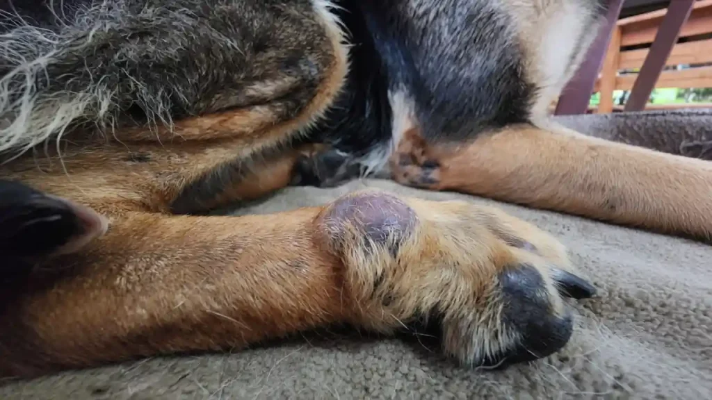
When you bring your Dog Skin Growth to the veterinarian for a skin growth, several diagnostic steps may be taken to determine its nature. The vet will first perform a thorough physical examination, noting the size, location, consistency, and mobility of the growth. However, visual inspection alone is rarely sufficient for a definitive diagnosis. More advanced diagnostics are usually required.
One of the most common initial diagnostic procedures is a Fine Needle Aspirate (FNA). During an FNA, a small, thin needle is inserted into the growth to collect a sample of cells. These cells are then spread on a slide and examined under a microscope by a veterinary pathologist (cytology). An FNA is quick, minimally invasive, and often can be performed without sedation, providing valuable information about the cell type and whether it appears benign or malignant.
If the FNA results are inconclusive, or if the vet suspects a more serious condition, a biopsy may be recommended. A biopsy involves removing a larger piece of tissue from the growth, which is then sent to a pathology lab for histological examination. Biopsies can be incisional (removing a part of the growth) or excisional (removing the entire growth). This procedure usually requires sedation or general anesthesia. The pathologist will analyze the tissue structure and cell characteristics to provide a definitive diagnosis, including the type of tumor, its grade (how aggressive it appears), and whether surgical margins are clear if the entire mass was removed.
In some cases, especially with suspected malignant growths, further diagnostics may be necessary to determine if the cancer has spread (staging). This can include blood tests, urinalysis, X-rays of the chest (to check for lung metastasis), and abdominal ultrasound. These tests help your veterinarian assess your dog’s overall health and develop the most appropriate treatment plan.
| Growth Type | Common Characteristics | Typical Location | Prognosis |
|---|---|---|---|
| Lipoma | Soft, movable, fatty, non-painful | Trunk, limbs, armpits | Excellent (benign) |
| Sebaceous Cyst | Firm, sometimes fluid-filled, may rupture | Head, neck, trunk | Good (benign, but can recur) |
| Histiocytoma | Rapidly growing, dome-shaped, red | Head, ears, limbs (young Dog Skin Growth ) | Excellent (benign, often self-resolving) |
| Skin Tag | Small, flesh-colored/pigmented, stalk-like | Areas of friction (armpits, groin) | Excellent (benign) |
| Mast Cell Tumor | Variable: small lump, red, itchy, ulcerated | Anywhere, including internal organs | Variable (malignant, can be aggressive) |
| Melanoma | Often dark, firm, can be non-pigmented | Mouth, nail beds, skin, eyes | Variable (malignant, especially oral/nail bed) |
| Squamous Cell Carcinoma | Raised, firm, ulcerated, may bleed | Sun-exposed areas (nose, ears, eyelids) | Fair to Guarded (malignant, locally invasive) |
Treatment Options for Canine Skin Growths

The treatment for a dog’s skin growth depends heavily on the diagnosis – whether it’s benign or malignant, its type, size, location, and the Dog Skin Growth overall health. Your veterinarian will discuss the best course of action once a definitive diagnosis has been made.
Surgical Removal
Surgical excision is the most common and often preferred treatment for many types of skin growths, both benign and malignant. For benign growths that are causing discomfort, irritation, or are simply cosmetically undesirable, surgical removal can be curative. For malignant tumors, surgery aims to remove the entire mass with wide, clear margins to prevent recurrence and spread. The success of surgical removal for malignant tumors depends on the type of cancer, its grade, and whether it has already metastasized. In some cases, if complete removal is not possible due to the tumor’s location or size, “debulking” surgery may be performed to reduce the tumor burden, followed by other treatments.
Non-Surgical Treatments
For certain types of growths, or when surgery is not an option due to the Dog Skin Growth age, health, or tumor location, non-surgical treatments may be considered:
- Chemotherapy: This involves using drugs to kill cancer cells throughout the body. It may be used as a standalone treatment for systemic cancers, or in conjunction with surgery or radiation for malignant skin tumors to target residual cancer cells or prevent metastasis.
- Radiation Therapy: This uses high-energy radiation to damage and kill cancer cells. It is often used for localized tumors that cannot be completely removed surgically or for tumors that are sensitive to radiation. It can be palliative (to reduce symptoms) or curative.
- Cryosurgery: This technique uses extreme cold (liquid nitrogen) to freeze and destroy abnormal tissue. It’s often used for small, superficial benign growths or certain types of early-stage skin cancers.
- Laser Therapy: Some growths can be removed using a medical laser, which can offer precise removal with less bleeding and pain.
- Topical Medications: For very superficial lesions, especially those caused by viruses (like papillomas), topical creams or ointments may sometimes be used.
- Observation: For certain benign growths, particularly lipomas that are not growing or causing problems, or histiocytomas in young Dog Skin Growth , a “watch and wait” approach may be recommended. Regular monitoring by the owner and periodic veterinary check-ups are essential for observing any changes.
The choice of treatment will be individualized based on the specific circumstances of each case, with your veterinarian guiding you through the decision-making process.
Preventing and Monitoring Skin Growths
While it’s impossible to completely prevent all skin growths in Dog Skin Growth , there are steps pet owners can take to minimize risks and ensure early detection, which is key to successful treatment, especially for malignant conditions.
Regular Home Checks
One of the most effective measures is to routinely perform full-body checks on your Dog Skin Growth . This involves gently running your hands over their entire body, paying close attention to areas that are often hidden by fur, such as the armpits, groin, ears, mouth, and paw pads. Feel for any new lumps, bumps, scabs, or changes in existing ones. Make this a positive experience for your dog, perhaps during grooming or cuddle time. A monthly check is a good habit to establish. If you find something, note its size, location, and date of discovery, and observe if it changes.
Diet and Lifestyle Factors
Maintaining your Dog Skin Growth optimal health through a balanced diet, regular exercise, and maintaining a healthy weight can contribute to overall well-being and potentially reduce the risk of certain health issues, including some types of growths. While direct causal links between diet and specific skin cancers are complex and not fully understood, a strong immune system supported by good nutrition is always beneficial. Protecting your Dog Skin Growth from excessive sun exposure, especially if they have short, light-colored fur, can help prevent squamous cell carcinoma and other sun-induced skin lesions.
Breed Predispositions
Awareness of breed-specific predispositions can also be helpful. Some breeds are more prone to certain types of skin growths. For instance, Boxers, Boston Terriers, and Pugs are genetically predisposed to mast cell tumors. Labrador Retrievers and Doberman Pinschers often develop lipomas. Knowing your Dog Skin Growth breed-specific risks can encourage more diligent monitoring and discussion with your vet about proactive screening measures.
Importance of Early Detection
Early detection is paramount, especially for malignant growths. The smaller a cancerous tumor is when detected and treated, the better the prognosis for complete remission or successful management. Regular veterinary check-ups, even for seemingly healthy Dog Skin Growth , are essential as vets can identify subtle changes that owners might miss. Professional grooming sessions can also sometimes reveal growths that owners haven’t noticed.
The Emotional Impact on Pet Owners
Discovering a skin growth on your Dog Skin Growth can be emotionally taxing. It’s natural to feel worried, anxious, or even guilty. Remember that many growths are benign, and even cancerous ones often have good treatment outcomes, particularly with early intervention. Lean on your veterinarian for support and clear communication, and don’t hesitate to seek a second opinion if you feel it’s necessary. Sharing your concerns with friends, family, or online pet owner communities can also provide emotional support.
The Role of Digital Photography in Monitoring
In today’s digital age, your smartphone can be a powerful tool in monitoring your dog’s skin growths. Taking clear, well-lit photographs of any new or existing lumps, along with a ruler or coin for scale, can create a visual record. Dating these photos allows you and your vet to track changes in size, color, and texture over time more accurately. This visual documentation can be invaluable in deciding whether a growth is stable, growing, or changing in a concerning way, aiding in the decision for further diagnostic steps.
Conclusion
Dog skin growth pictures serve as an initial point of reference for many pet owners, but they are just the beginning of understanding these canine health concerns. While a visual guide can offer preliminary insights, it is absolutely critical to remember that accurate diagnosis and appropriate treatment for any skin growth can only be provided by a qualified veterinarian. From benign lipomas to aggressive mast cell tumors, the range of possible growths is vast, and their implications for your Dog Skin Growthhealth vary significantly. Regular physical examinations at home, coupled with routine veterinary check-ups, are your best tools for early detection. By being vigilant and proactive, you can ensure that any suspicious lump or bump on your beloved canine companion receives the timely attention it needs, ultimately safeguarding their health and well-being.